7KBR

Co-crystal structure of alpha glucosidase with compound 10


Experimental Data Snapshot

  • Method: X-RAY DIFFRACTION
  • Resolution: 2.09 Å
  • R-Value Free: 0.186 
  • R-Value Work: 0.163 
  • R-Value Observed: 0.164 

Starting Model: experimental
View more details

wwPDB Validation   3D Report Full Report


Ligand Structure Quality Assessment 


This is version 1.4 of the entry. See complete history


Literature

N-Substituted Valiolamine Derivatives as Potent Inhibitors of Endoplasmic Reticulum alpha-Glucosidases I and II with Antiviral Activity.

Karade, S.S.Hill, M.L.Kiappes, J.L.Manne, R.Aakula, B.Zitzmann, N.Warfield, K.L.Treston, A.M.Mariuzza, R.A.

(2021) J Med Chem 64: 18010-18024

  • DOI: https://doi.org/10.1021/acs.jmedchem.1c01377
  • Primary Citation of Related Structures:  
    7JTY, 7K9N, 7K9O, 7K9Q, 7K9T, 7KAD, 7KB6, 7KB8, 7KBJ, 7KBR, 7KRY, 7L9E

  • PubMed Abstract: 

    Most enveloped viruses rely on the host cell endoplasmic reticulum (ER) quality control (QC) machinery for proper folding of glycoproteins. The key ER α-glucosidases (α-Glu) I and II of the ERQC machinery are attractive targets for developing broad-spectrum antivirals. Iminosugars based on deoxynojirimycin have been extensively studied as ER α-glucosidase inhibitors; however, other glycomimetic compounds are less established. Accordingly, we synthesized a series of N-substituted derivatives of valiolamine, the iminosugar scaffold of type 2 diabetes drug voglibose. To understand the basis for up to 100,000-fold improved inhibitory potency, we determined high-resolution crystal structures of mouse ER α-GluII in complex with valiolamine and 10 derivatives. The structures revealed extensive interactions with all four α-GluII subsites. We further showed that N-substituted valiolamines were active against dengue virus and SARS-CoV-2 in vitro . This study introduces valiolamine-based inhibitors of the ERQC machinery as candidates for developing potential broad-spectrum therapeutics against the existing and emerging viruses.


  • Organizational Affiliation

    University of Maryland Institute for Bioscience and Biotechnology Research, Rockville, Maryland 20850, United States.


Macromolecules
Find similar proteins by:  (by identity cutoff)  |  3D Structure
Entity ID: 1
MoleculeChains Sequence LengthOrganismDetailsImage
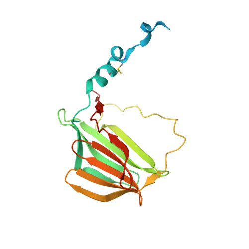
Neutral alpha-glucosidase AB Trypsin-cleaved Fragment #1A [auth G],
E [auth I]
183Mus musculusMutation(s): 1 
Gene Names: GanabG2anKiaa0088
EC: 3.2.1.207
UniProt
Find proteins for Q8BHN3 (Mus musculus)
Explore Q8BHN3 
Go to UniProtKB:  Q8BHN3
Entity Groups  
Sequence Clusters30% Identity50% Identity70% Identity90% Identity95% Identity100% Identity
UniProt GroupQ8BHN3
Sequence Annotations
Expand
  • Reference Sequence
Find similar proteins by:  (by identity cutoff)  |  3D Structure
Entity ID: 2
MoleculeChains Sequence LengthOrganismDetailsImage
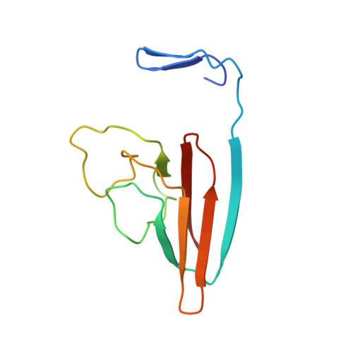
Neutral alpha-glucosidase AB Trypsin-cleaved Fragment #2B [auth H],
F [auth J]
107Mus musculusMutation(s): 0 
Gene Names: GanabG2anKiaa0088
EC: 3.2.1.207
UniProt
Find proteins for Q8BHN3 (Mus musculus)
Explore Q8BHN3 
Go to UniProtKB:  Q8BHN3
Entity Groups  
Sequence Clusters30% Identity50% Identity70% Identity90% Identity95% Identity100% Identity
UniProt GroupQ8BHN3
Sequence Annotations
Expand
  • Reference Sequence
Find similar proteins by:  (by identity cutoff)  |  3D Structure
Entity ID: 3
MoleculeChains Sequence LengthOrganismDetailsImage
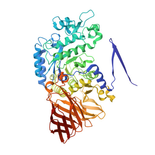
Neutral alpha-glucosidase AB Trypsin-cleaved Fragment #3C [auth A],
G [auth C]
609Mus musculusMutation(s): 0 
Gene Names: GanabG2anKiaa0088
EC: 3.2.1.207
UniProt
Find proteins for Q8BHN3 (Mus musculus)
Explore Q8BHN3 
Go to UniProtKB:  Q8BHN3
Entity Groups  
Sequence Clusters30% Identity50% Identity70% Identity90% Identity95% Identity100% Identity
UniProt GroupQ8BHN3
Sequence Annotations
Expand
  • Reference Sequence
Find similar proteins by:  (by identity cutoff)  |  3D Structure
Entity ID: 4
MoleculeChains Sequence LengthOrganismDetailsImage
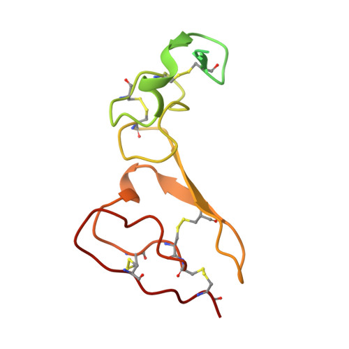
Glucosidase 2 subunit betaD [auth B],
H [auth D]
134Mus musculusMutation(s): 0 
Gene Names: Prkcsh
UniProt & NIH Common Fund Data Resources
Find proteins for O08795 (Mus musculus)
Explore O08795 
Go to UniProtKB:  O08795
IMPC:  MGI:107877
Entity Groups  
Sequence Clusters30% Identity50% Identity70% Identity90% Identity95% Identity100% Identity
UniProt GroupO08795
Sequence Annotations
Expand
  • Reference Sequence
Small Molecules
Ligands 7 Unique
IDChains Name / Formula / InChI Key2D Diagram3D Interactions
WAS (Subject of Investigation/LOI)
Query on WAS

Download Ideal Coordinates CCD File 
IA [auth A],
QB [auth C]
2-{[2-nitro-4-(triazan-1-yl)phenyl]amino}ethyl (2-{[(1S,2S,3R,4S,5S)-2,3,4,5-tetrahydroxy-5-(hydroxymethyl)cyclohexyl]amino}ethyl)carbamate
C18 H31 N7 O9
RDKDXFWNGAUKLV-IJCAJFLPSA-N
PG4
Query on PG4

Download Ideal Coordinates CCD File 
U [auth A],
UB [auth D],
ZA [auth C]
TETRAETHYLENE GLYCOL
C8 H18 O5
UWHCKJMYHZGTIT-UHFFFAOYSA-N
PGE
Query on PGE

Download Ideal Coordinates CCD File 
KA [auth A],
Q [auth A],
QA [auth B],
T [auth A],
Y [auth A]
TRIETHYLENE GLYCOL
C6 H14 O4
ZIBGPFATKBEMQZ-UHFFFAOYSA-N
PEG
Query on PEG

Download Ideal Coordinates CCD File 
AB [auth C]
BA [auth A]
DA [auth A]
EA [auth A]
GB [auth C]
AB [auth C],
BA [auth A],
DA [auth A],
EA [auth A],
GB [auth C],
HA [auth A],
HB [auth C],
K [auth G],
KB [auth C],
LB [auth C],
OB [auth C],
V [auth A],
XA [auth C],
Z [auth A]
DI(HYDROXYETHYL)ETHER
C4 H10 O3
MTHSVFCYNBDYFN-UHFFFAOYSA-N
SO4
Query on SO4

Download Ideal Coordinates CCD File 
L [auth G]
LA [auth A]
MA [auth A]
N [auth H]
NA [auth A]
L [auth G],
LA [auth A],
MA [auth A],
N [auth H],
NA [auth A],
RB [auth C],
SB [auth C],
TA [auth B]
SULFATE ION
O4 S
QAOWNCQODCNURD-UHFFFAOYSA-L
EDO
Query on EDO

Download Ideal Coordinates CCD File 
AA [auth A]
BB [auth C]
CA [auth A]
CB [auth C]
DB [auth C]
AA [auth A],
BB [auth C],
CA [auth A],
CB [auth C],
DB [auth C],
EB [auth C],
FA [auth A],
FB [auth C],
GA [auth A],
I [auth G],
IB [auth C],
J [auth G],
JA [auth A],
JB [auth C],
M [auth H],
MB [auth C],
NB [auth C],
O [auth A],
OA [auth B],
P [auth A],
PA [auth B],
PB [auth C],
R [auth A],
S [auth A],
TB [auth D],
UA [auth I],
VA [auth J],
W [auth A],
WA [auth J],
X [auth A],
YA [auth C]
1,2-ETHANEDIOL
C2 H6 O2
LYCAIKOWRPUZTN-UHFFFAOYSA-N
CA
Query on CA

Download Ideal Coordinates CCD File 
RA [auth B],
SA [auth B],
VB [auth D],
WB [auth D]
CALCIUM ION
Ca
BHPQYMZQTOCNFJ-UHFFFAOYSA-N
Experimental Data & Validation

Experimental Data

  • Method: X-RAY DIFFRACTION
  • Resolution: 2.09 Å
  • R-Value Free: 0.186 
  • R-Value Work: 0.163 
  • R-Value Observed: 0.164 
  • Space Group: P 32
Unit Cell:
Length ( Å )Angle ( ˚ )
a = 102.815α = 90
b = 102.815β = 90
c = 240.552γ = 120
Software Package:
Software NamePurpose
PHENIXrefinement
PDB_EXTRACTdata extraction
HKL-2000data reduction
HKL-2000data scaling
PHENIXphasing

Structure Validation

View Full Validation Report


Ligand Structure Quality Assessment 


Entry History 

Deposition Data

Revision History  (Full details and data files)

  • Version 1.0: 2021-10-06
    Type: Initial release
  • Version 1.1: 2021-12-22
    Changes: Database references
  • Version 1.2: 2022-01-05
    Changes: Database references
  • Version 1.3: 2023-10-18
    Changes: Data collection, Refinement description
  • Version 1.4: 2024-12-25
    Changes: Advisory, Derived calculations, Structure summary