7KBJ | pdb_00007kbj

Co-crystal structure of alpha glucosidase with compound 9


Experimental Data Snapshot

  • Method: X-RAY DIFFRACTION
  • Resolution: 2.21 Å
  • R-Value Free: 
    0.200 (Depositor), 0.199 (DCC) 
  • R-Value Work: 
    0.167 (Depositor), 0.167 (DCC) 
  • R-Value Observed: 
    0.167 (Depositor) 

Starting Model: experimental
View more details

wwPDB Validation   3D Report Full Report


Ligand Structure Quality Assessment 

Created with Raphaël 2.3.0Worse 01 BetterLigand structure goodness of fit to experimental dataBest fitted WAVClick on this verticalbar to view details

This is version 1.4 of the entry. See complete history


Literature

N-Substituted Valiolamine Derivatives as Potent Inhibitors of Endoplasmic Reticulum alpha-Glucosidases I and II with Antiviral Activity.

Karade, S.S.Hill, M.L.Kiappes, J.L.Manne, R.Aakula, B.Zitzmann, N.Warfield, K.L.Treston, A.M.Mariuzza, R.A.

(2021) J Med Chem 64: 18010-18024

  • DOI: https://doi.org/10.1021/acs.jmedchem.1c01377
  • Primary Citation of Related Structures:  
    7JTY, 7K9N, 7K9O, 7K9Q, 7K9T, 7KAD, 7KB6, 7KB8, 7KBJ, 7KBR, 7KRY, 7L9E

  • PubMed Abstract: 

    Most enveloped viruses rely on the host cell endoplasmic reticulum (ER) quality control (QC) machinery for proper folding of glycoproteins. The key ER α-glucosidases (α-Glu) I and II of the ERQC machinery are attractive targets for developing broad-spectrum antivirals. Iminosugars based on deoxynojirimycin have been extensively studied as ER α-glucosidase inhibitors; however, other glycomimetic compounds are less established. Accordingly, we synthesized a series of N-substituted derivatives of valiolamine, the iminosugar scaffold of type 2 diabetes drug voglibose. To understand the basis for up to 100,000-fold improved inhibitory potency, we determined high-resolution crystal structures of mouse ER α-GluII in complex with valiolamine and 10 derivatives. The structures revealed extensive interactions with all four α-GluII subsites. We further showed that N-substituted valiolamines were active against dengue virus and SARS-CoV-2 in vitro . This study introduces valiolamine-based inhibitors of the ERQC machinery as candidates for developing potential broad-spectrum therapeutics against the existing and emerging viruses.


  • Organizational Affiliation

    University of Maryland Institute for Bioscience and Biotechnology Research, Rockville, Maryland 20850, United States.


Macromolecules
Find similar proteins by:  (by identity cutoff)  |  3D Structure
Entity ID: 1
MoleculeChains Sequence LengthOrganismDetailsImage
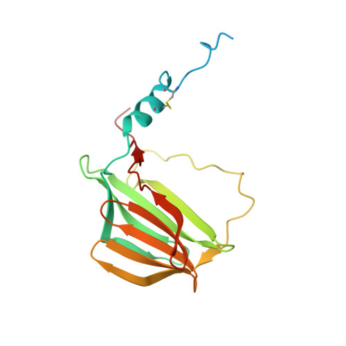
Neutral alpha-glucosidase AB Trypsin-cleaved Fragment #1A [auth G],
E [auth I]
184Mus musculusMutation(s): 1 
Gene Names: GanabG2anKiaa0088
EC: 3.2.1.207
UniProt
Find proteins for Q8BHN3 (Mus musculus)
Explore Q8BHN3 
Go to UniProtKB:  Q8BHN3
Entity Groups  
Sequence Clusters30% Identity50% Identity70% Identity90% Identity95% Identity100% Identity
UniProt GroupQ8BHN3
Sequence Annotations
Expand
  • Reference Sequence
Find similar proteins by:  (by identity cutoff)  |  3D Structure
Entity ID: 2
MoleculeChains Sequence LengthOrganismDetailsImage
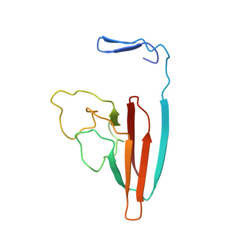
Neutral alpha-glucosidase AB Trypsin-cleaved Fragment #2B [auth H],
F [auth J]
107Mus musculusMutation(s): 0 
Gene Names: GanabG2anKiaa0088
EC: 3.2.1.207
UniProt
Find proteins for Q8BHN3 (Mus musculus)
Explore Q8BHN3 
Go to UniProtKB:  Q8BHN3
Entity Groups  
Sequence Clusters30% Identity50% Identity70% Identity90% Identity95% Identity100% Identity
UniProt GroupQ8BHN3
Sequence Annotations
Expand
  • Reference Sequence
Find similar proteins by:  (by identity cutoff)  |  3D Structure
Entity ID: 3
MoleculeChains Sequence LengthOrganismDetailsImage
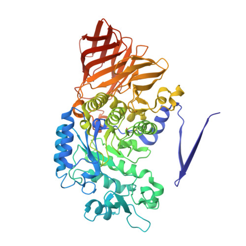
Neutral alpha-glucosidase AB Trypsin-cleaved Fragment #3C [auth A],
G [auth C]
609Mus musculusMutation(s): 0 
Gene Names: GanabG2anKiaa0088
EC: 3.2.1.207
UniProt
Find proteins for Q8BHN3 (Mus musculus)
Explore Q8BHN3 
Go to UniProtKB:  Q8BHN3
Entity Groups  
Sequence Clusters30% Identity50% Identity70% Identity90% Identity95% Identity100% Identity
UniProt GroupQ8BHN3
Sequence Annotations
Expand
  • Reference Sequence
Find similar proteins by:  (by identity cutoff)  |  3D Structure
Entity ID: 4
MoleculeChains Sequence LengthOrganismDetailsImage
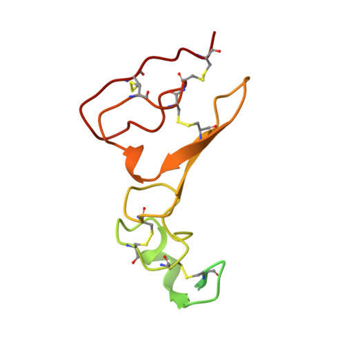
Glucosidase 2 subunit betaD [auth B],
H [auth D]
134Mus musculusMutation(s): 0 
Gene Names: Prkcsh
UniProt & NIH Common Fund Data Resources
Find proteins for O08795 (Mus musculus)
Explore O08795 
Go to UniProtKB:  O08795
IMPC:  MGI:107877
Entity Groups  
Sequence Clusters30% Identity50% Identity70% Identity90% Identity95% Identity100% Identity
UniProt GroupO08795
Sequence Annotations
Expand
  • Reference Sequence
Small Molecules
Ligands 7 Unique
IDChains Name / Formula / InChI Key2D Diagram3D Interactions
WAV (Subject of Investigation/LOI)
Query on WAV

Download Ideal Coordinates CCD File 
O [auth A],
TA [auth C]
(1S,2S,3R,4S,5S)-1-(hydroxymethyl)-5-{[2-(2-{[2-nitro-4-(triazan-1-yl)phenyl]amino}ethoxy)ethyl]amino}cyclohexane-1,2,3,4-tetrol
C17 H30 N6 O8
DYANQIPKBSETSL-RKOFLFEYSA-N
PG4
Query on PG4

Download Ideal Coordinates CCD File 
L [auth H],
P [auth A]
TETRAETHYLENE GLYCOL
C8 H18 O5
UWHCKJMYHZGTIT-UHFFFAOYSA-N
PGE
Query on PGE

Download Ideal Coordinates CCD File 
I [auth G],
IA [auth A],
Q [auth A],
U [auth A],
V [auth A]
TRIETHYLENE GLYCOL
C6 H14 O4
ZIBGPFATKBEMQZ-UHFFFAOYSA-N
PEG
Query on PEG

Download Ideal Coordinates CCD File 
BA [auth A]
IB [auth C]
KA [auth B]
LA [auth B]
OB [auth C]
DI(HYDROXYETHYL)ETHER
C4 H10 O3
MTHSVFCYNBDYFN-UHFFFAOYSA-N
SO4
Query on SO4

Download Ideal Coordinates CCD File 
JA [auth A]
K [auth G]
MA [auth B]
PB [auth C]
QB [auth C]
SULFATE ION
O4 S
QAOWNCQODCNURD-UHFFFAOYSA-L
EDO
Query on EDO

Download Ideal Coordinates CCD File 
AA [auth A]
AB [auth C]
BB [auth C]
CA [auth A]
CB [auth C]
1,2-ETHANEDIOL
C2 H6 O2
LYCAIKOWRPUZTN-UHFFFAOYSA-N
CA
Query on CA

Download Ideal Coordinates CCD File 
NA [auth B],
OA [auth B],
VB [auth D],
WB [auth D]
CALCIUM ION
Ca
BHPQYMZQTOCNFJ-UHFFFAOYSA-N
Experimental Data & Validation

Experimental Data

  • Method: X-RAY DIFFRACTION
  • Resolution: 2.21 Å
  • R-Value Free:  0.200 (Depositor), 0.199 (DCC) 
  • R-Value Work:  0.167 (Depositor), 0.167 (DCC) 
  • R-Value Observed: 0.167 (Depositor) 
Space Group: P 32
Unit Cell:
Length ( Å )Angle ( ˚ )
a = 102.652α = 90
b = 102.652β = 90
c = 238.908γ = 120
Software Package:
Software NamePurpose
PHENIXrefinement
PDB_EXTRACTdata extraction
HKL-2000data reduction
HKL-2000data scaling
PHENIXphasing

Structure Validation

View Full Validation Report


Ligand Structure Quality Assessment 

Created with Raphaël 2.3.0Worse 01 BetterLigand structure goodness of fit to experimental dataBest fitted WAVClick on this verticalbar to view details

Entry History 

Deposition Data

Revision History  (Full details and data files)

  • Version 1.0: 2021-10-06
    Type: Initial release
  • Version 1.1: 2021-12-22
    Changes: Database references
  • Version 1.2: 2022-01-05
    Changes: Database references
  • Version 1.3: 2023-10-18
    Changes: Data collection, Refinement description
  • Version 1.4: 2024-12-25
    Changes: Advisory, Derived calculations, Structure summary