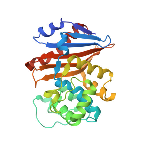
7KHQ

Crystal structure of OXA-48 K73A in complex with meropenem


Experimental Data Snapshot

  • Method: X-RAY DIFFRACTION
  • Resolution: 2.00 Å
  • R-Value Free: 0.236 
  • R-Value Work: 0.207 
  • R-Value Observed: 0.209 

Starting Model: experimental
View more details

wwPDB Validation   3D Report Full Report


Ligand Structure Quality Assessment 


This is version 1.4 of the entry. See complete history


Literature

Mechanistic Basis of OXA-48-like beta-Lactamases' Hydrolysis of Carbapenems.

Stojanoski, V.Hu, L.Sankaran, B.Wang, F.Tao, P.Prasad, B.V.V.Palzkill, T.

(2021) ACS Infect Dis 7: 445-460

  • DOI: https://doi.org/10.1021/acsinfecdis.0c00798
  • Primary Citation of Related Structures:  
    7KH9, 7KHQ, 7KHY, 7KHZ

  • PubMed Abstract: 

    Carbapenem-hydrolyzing class D β-lactamases (CHDLs) are an important source of resistance to these last resort β-lactam antibiotics. OXA-48 is a member of a group of CHDLs named OXA-48-like enzymes. On the basis of sequence similarity, OXA-163 can be classified as an OXA-48-like enzyme, but it has altered substrate specificity. Compared to OXA-48, it shows impaired activity for carbapenems but displays an enhanced hydrolysis of oxyimino-cephalosporins. Here, we address the mechanistic and structural basis for carbapenem hydrolysis by OXA-48-like enzymes. Pre-steady-state kinetic analysis indicates that the rate-limiting step for OXA-48 and OXA-163 hydrolysis of carbapenems is deacylation and that the greatly reduced carbapenemase activity of OXA-163 compared to that of OXA-48 is due entirely to a slower deacylation reaction. Furthermore, our structural data indicate that the positioning of the β5-β6 loop is necessary for carbapenem hydrolysis by OXA-48. A major difference between the OXA-48 and OXA-163 complexes with carbapenems is that the 214-RIEP-217 deletion in OXA-163 creates a large opening in the active site that is absent in the OXA-48/carbapenem structures. We propose that the larger active site results in less constraint on the conformation of the 6α-hydroxyethyl group in the acyl-enzyme. The acyl-enzyme intermediate assumes multiple conformations, most of which are incompatible with rapid deacylation. Consistent with this hypothesis, molecular dynamics simulations indicate that the most stable complex is formed between OXA-48 and imipenem, which correlates with the OXA-48 hydrolysis of imipenem being the fastest observed. Furthermore, the OXA-163 complexes with imipenem and meropenem are the least stable and show significant conformational fluctuations, which correlates with the slow hydrolysis of these substrates.


  • Organizational Affiliation

    Department of Molecular Biophysics and Integrated Bioimaging, Berkeley Center for Structural Biology, Lawrence Berkeley National Laboratory, Berkeley, California 94720, United States.


Macromolecules
Find similar proteins by:  (by identity cutoff)  |  3D Structure
Entity ID: 1
MoleculeChains Sequence LengthOrganismDetailsImage
Beta-lactamase
A, B
244Klebsiella pneumoniaeMutation(s): 1 
Gene Names: 
EC: 3.5.2.6
UniProt
Find proteins for Q6XEC0 (Klebsiella pneumoniae)
Explore Q6XEC0 
Go to UniProtKB:  Q6XEC0
Entity Groups  
Sequence Clusters30% Identity50% Identity70% Identity90% Identity95% Identity100% Identity
UniProt GroupQ6XEC0
Sequence Annotations
Expand
  • Reference Sequence
Small Molecules
Ligands 3 Unique
IDChains Name / Formula / InChI Key2D Diagram3D Interactions
MER (Subject of Investigation/LOI)
Query on MER

Download Ideal Coordinates CCD File 
C [auth A],
G [auth B]
(4R,5S)-3-{[(3S,5S)-5-(dimethylcarbamoyl)pyrrolidin-3-yl]sulfanyl}-5-[(2S,3R)-3-hydroxy-1-oxobutan-2-yl]-4-methyl-4,5-d ihydro-1H-pyrrole-2-carboxylic acid
C17 H27 N3 O5 S
DYQHXZPAIVAJRU-HTXLXMOSSA-N
ZN
Query on ZN

Download Ideal Coordinates CCD File 
F [auth A],
J [auth B]
ZINC ION
Zn
PTFCDOFLOPIGGS-UHFFFAOYSA-N
CL
Query on CL

Download Ideal Coordinates CCD File 
D [auth A],
E [auth A],
H [auth B],
I [auth B]
CHLORIDE ION
Cl
VEXZGXHMUGYJMC-UHFFFAOYSA-M
Experimental Data & Validation

Experimental Data

  • Method: X-RAY DIFFRACTION
  • Resolution: 2.00 Å
  • R-Value Free: 0.236 
  • R-Value Work: 0.207 
  • R-Value Observed: 0.209 
  • Space Group: P 2 21 21
Unit Cell:
Length ( Å )Angle ( ˚ )
a = 44.285α = 90
b = 105.487β = 90
c = 125.665γ = 90
Software Package:
Software NamePurpose
PHENIXrefinement
DIALSdata reduction
DIALSdata scaling
MOLREPphasing

Structure Validation

View Full Validation Report


Ligand Structure Quality Assessment 


Entry History & Funding Information

Deposition Data


Funding OrganizationLocationGrant Number
National Institutes of Health/National Institute Of Allergy and Infectious Diseases (NIH/NIAID)United States--

Revision History  (Full details and data files)

  • Version 1.0: 2021-02-10
    Type: Initial release
  • Version 1.1: 2021-02-24
    Changes: Database references
  • Version 1.2: 2021-06-30
    Changes: Structure summary
  • Version 1.3: 2023-10-18
    Changes: Data collection, Database references, Refinement description
  • Version 1.4: 2024-11-06
    Changes: Structure summary