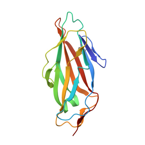
8BXY | pdb_00008bxy

FimH in complex with alpha1,6 core-fucosylated oligomannose-3, crystallized in the trigonal space group


Experimental Data Snapshot

  • Method: X-RAY DIFFRACTION
  • Resolution: 1.45 Å
  • R-Value Free: 
    0.205 (Depositor), 0.200 (DCC) 
  • R-Value Work: 
    0.153 (Depositor), 0.150 (DCC) 

Starting Model: experimental
View more details

wwPDB Validation   3D Report Full Report


Ligand Structure Quality Assessment 

Created with Raphaël 2.3.0Worse 01 BetterLigand structure goodness of fit to experimental dataBest fitted SO4Click on this verticalbar to view details

This is version 1.3 of the entry. See complete history

Re-refinement Note

This entry reflects an alternative modeling of the original data in: 7BHD


Literature

Structural insights into a cooperative switch between one and two FimH bacterial adhesins binding pauci- and high-mannose type N-glycan receptors.

Krammer, E.M.Bridot, C.Serna, S.Echeverria, B.Semwal, S.Roubinet, B.van Noort, K.Wilbers, R.H.P.Bourenkov, G.de Ruyck, J.Landemarre, L.Reichardt, N.Bouckaert, J.

(2023) J Biological Chem 299: 104627-104627

  • DOI: https://doi.org/10.1016/j.jbc.2023.104627
  • Primary Citation of Related Structures:  
    7BHD, 8BXY, 8BY3

  • PubMed Abstract: 

    The FimH type-1 fimbrial adhesin allows pathogenic Escherichia coli to adhere to glycoproteins in the epithelial linings of human bladder and intestinal tract, by using multiple fimbriae simultaneously. Pauci- and high-mannose type N-glycans are natural FimH receptors on those glycoproteins. Oligomannose-3 and oligomannose-5 bind with the highest affinity to FimH by using the same Manα1,3Man branch. Oligomannose-6 is generated from oligomannose-5 in the next step of the biogenesis of high-mannose N-glycans, by the transfer of a mannose in α1,2-linkage onto this branch. Using serial crystallography and by measuring the kinetics of binding, we demonstrate that shielding the high-affinity epitope drives the binding of multiple FimH molecules. First, we profiled FimH glycan binding on a microarray containing paucimannosidic N-glycans and in a FimH LEctPROFILE assay. To make the transition to oligomannose-6, we measured the kinetics of FimH binding using paucimannosidic N-glycans, glycoproteins and all four α-dimannosides conjugated to bovine serum albumin. Equimolar mixed interfaces of the dimannosides present in oligomannose-6 and molecular dynamics simulations suggest a positive cooperativity in the bivalent binding of Manα1,3Manα1 and Manα1,6Manα1 dimannosides. The binding of core α1,6-fucosylated oligomannose-3 in cocrystals of FimH is monovalent but interestingly the GlcNAc1-Fuc moiety retains highly flexibility. In cocrystals with oligomannose-6, two FimH bacterial adhesins bind the Manα1,3Manα1 and Manα1,6Manα1 endings of the second trimannose core (A-4'-B). This cooperative switch towards bivalent binding appears sustainable beyond a molar excess of oligomannose-6. Our findings provide important novel structural insights for the design of multivalent FimH antagonists that bind with positive cooperativity.


  • Organizational Affiliation

    Unité de Glycobiologie Structurale et Fonctionnelle (UGSF), UMR 8576 CNRS and University of Lille, Villeneuve d'Ascq, France.


Macromolecules
Find similar proteins by:  (by identity cutoff)  |  3D Structure
Entity ID: 1
MoleculeChains Sequence LengthOrganismDetailsImage
Type 1 fimbrin D-mannose specific adhesin
A, B
158Escherichia coli K-12Mutation(s): 0 
Gene Names: fimHb4320JW4283
UniProt
Find proteins for P08191 (Escherichia coli (strain K12))
Explore P08191 
Go to UniProtKB:  P08191
Entity Groups  
Sequence Clusters30% Identity50% Identity70% Identity90% Identity95% Identity100% Identity
UniProt GroupP08191
Sequence Annotations
Expand
  • Reference Sequence
Oligosaccharides

Help

Entity ID: 2
MoleculeChains Length2D Diagram Glycosylation3D Interactions
alpha-D-mannopyranose-(1-3)-[alpha-D-mannopyranose-(1-6)]beta-D-mannopyranose-(1-4)-2-acetamido-2-deoxy-beta-D-glucopyranose-(1-4)-[alpha-L-fucopyranose-(1-6)]2-acetamido-2-deoxy-beta-D-glucopyranose
C, D
6N/A
Glycosylation Resources
GlyTouCan:  G82348BZ
GlyCosmos:  G82348BZ
GlyGen:  G82348BZ
Experimental Data & Validation

Experimental Data

  • Method: X-RAY DIFFRACTION
  • Resolution: 1.45 Å
  • R-Value Free:  0.205 (Depositor), 0.200 (DCC) 
  • R-Value Work:  0.153 (Depositor), 0.150 (DCC) 
Space Group: P 31 2 1
Unit Cell:
Length ( Å )Angle ( ˚ )
a = 91.056α = 90
b = 91.056β = 90
c = 79.578γ = 120
Software Package:
Software NamePurpose
REFMACrefinement
PHENIXrefinement
PHENIXmodel building
autoPROCdata processing
XDSdata reduction
XSCALEdata scaling
PHASERphasing

Structure Validation

View Full Validation Report


Ligand Structure Quality Assessment 

Created with Raphaël 2.3.0Worse 01 BetterLigand structure goodness of fit to experimental dataBest fitted SO4Click on this verticalbar to view details

Entry History & Funding Information

Deposition Data


Funding OrganizationLocationGrant Number
H2020 Marie Curie Actions of the European CommissionEuropean Union750280

Revision History  (Full details and data files)

  • Version 1.0: 2023-04-12
    Type: Initial release
  • Version 1.1: 2023-04-19
    Changes: Database references
  • Version 1.2: 2023-04-26
    Changes: Database references
  • Version 1.3: 2024-10-23
    Changes: Data collection, Refinement description, Structure summary