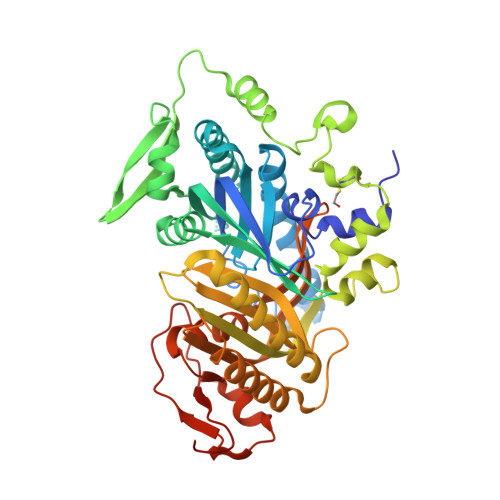
8COJ

Crystal structure of human soluble adenylyl cyclase catalytic domain in complex with the inhibitor TDI-10228


Experimental Data Snapshot

  • Method: X-RAY DIFFRACTION
  • Resolution: 2.10 Å
  • R-Value Free: 0.225 
  • R-Value Work: 0.201 
  • R-Value Observed: 0.202 

Starting Model: experimental
View more details

wwPDB Validation   3D Report Full Report


Ligand Structure Quality Assessment 


This is version 1.2 of the entry. See complete history


Literature

Scaffold Hopping and Optimization of Small Molecule Soluble Adenyl Cyclase Inhibitors Led by Free Energy Perturbation.

Sun, S.Fushimi, M.Rossetti, T.Kaur, N.Ferreira, J.Miller, M.Quast, J.van den Heuvel, J.Steegborn, C.Levin, L.R.Buck, J.Myers, R.W.Kargman, S.Liverton, N.Meinke, P.T.Huggins, D.J.

(2023) J Chem Inf Model 63: 2828-2841

  • DOI: https://doi.org/10.1021/acs.jcim.2c01577
  • Primary Citation of Related Structures:  
    8CNH, 8CO7, 8COJ, 8COT

  • PubMed Abstract: 

    Free energy perturbation is a computational technique that can be used to predict how small changes to an inhibitor structure will affect the binding free energy to its target. In this paper, we describe the utility of free energy perturbation with FEP+ in the hit-to-lead stage of a drug discovery project targeting soluble adenyl cyclase. The project was structurally enabled by X-ray crystallography throughout. We employed free energy perturbation to first scaffold hop to a preferable chemotype and then optimize the binding affinity to sub-nanomolar levels while retaining druglike properties. The results illustrate that effective use of free energy perturbation can enable a drug discovery campaign to progress rapidly from hit to lead, facilitating proof-of-concept studies that enable target validation.


  • Organizational Affiliation

    Tri-Institutional Therapeutics Discovery Institute, New York, New York 10021, United States.


Macromolecules
Find similar proteins by:  (by identity cutoff)  |  3D Structure
Entity ID: 1
MoleculeChains Sequence LengthOrganismDetailsImage
Adenylate cyclase type 10475Homo sapiensMutation(s): 0 
Gene Names: ADCY10SAC
EC: 4.6.1.1
UniProt & NIH Common Fund Data Resources
Find proteins for Q96PN6 (Homo sapiens)
Explore Q96PN6 
Go to UniProtKB:  Q96PN6
PHAROS:  Q96PN6
GTEx:  ENSG00000143199 
Entity Groups  
Sequence Clusters30% Identity50% Identity70% Identity90% Identity95% Identity100% Identity
UniProt GroupQ96PN6
Sequence Annotations
Expand
  • Reference Sequence
Small Molecules
Ligands 4 Unique
IDChains Name / Formula / InChI Key2D Diagram3D Interactions
VE1 (Subject of Investigation/LOI)
Query on VE1

Download Ideal Coordinates CCD File 
B [auth A]4-chloranyl-6-[4-[(3-fluorophenyl)methyl]-1-methyl-pyrazol-3-yl]pyrimidin-2-amine
C15 H13 Cl F N5
NVFOMYRKSZVYGO-UHFFFAOYSA-N
DMS
Query on DMS

Download Ideal Coordinates CCD File 
C [auth A],
D [auth A],
E [auth A],
F [auth A]
DIMETHYL SULFOXIDE
C2 H6 O S
IAZDPXIOMUYVGZ-UHFFFAOYSA-N
EDO
Query on EDO

Download Ideal Coordinates CCD File 
H [auth A]
I [auth A]
J [auth A]
K [auth A]
L [auth A]
H [auth A],
I [auth A],
J [auth A],
K [auth A],
L [auth A],
M [auth A]
1,2-ETHANEDIOL
C2 H6 O2
LYCAIKOWRPUZTN-UHFFFAOYSA-N
ACT
Query on ACT

Download Ideal Coordinates CCD File 
G [auth A]ACETATE ION
C2 H3 O2
QTBSBXVTEAMEQO-UHFFFAOYSA-M
Modified Residues  1 Unique
IDChains TypeFormula2D DiagramParent
CME
Query on CME
A
L-PEPTIDE LINKINGC5 H11 N O3 S2CYS
Experimental Data & Validation

Experimental Data

  • Method: X-RAY DIFFRACTION
  • Resolution: 2.10 Å
  • R-Value Free: 0.225 
  • R-Value Work: 0.201 
  • R-Value Observed: 0.202 
  • Space Group: P 63
Unit Cell:
Length ( Å )Angle ( ˚ )
a = 98.905α = 90
b = 98.905β = 90
c = 99.746γ = 120
Software Package:
Software NamePurpose
PHENIXrefinement
XDSdata reduction
XDSdata scaling
PHASERphasing

Structure Validation

View Full Validation Report


Ligand Structure Quality Assessment 


Entry History & Funding Information

Deposition Data


Funding OrganizationLocationGrant Number
German Research Foundation (DFG)GermanySTE1701/11

Revision History  (Full details and data files)

  • Version 1.0: 2023-04-26
    Type: Initial release
  • Version 1.1: 2023-05-17
    Changes: Database references
  • Version 1.2: 2024-10-16
    Changes: Data collection, Structure summary