


X-ray crystal structure of Aggregatibacter actinomycetemcomitans dimanganese(II) class Id ribonucleotide reductase beta subunit
Rose, H.R., Boal, A.K.To be published.
Experimental Data Snapshot
Starting Model: experimental
View more details
wwPDB Validation 3D Report Full Report

Entity ID: 1 | |||||
|---|---|---|---|---|---|
| Molecule | Chains | Sequence Length | Organism | Details | Image |
| Ribonucleoside-diphosphate reductase | 348 | Aggregatibacter actinomycetemcomitans | Mutation(s): 0 Gene Names: FXB68_03595 EC: 1.17.4.1 | 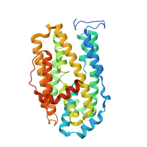 | |
UniProt | |||||
Find proteins for A0A142G3G1 (Aggregatibacter actinomycetemcomitans) Explore A0A142G3G1 Go to UniProtKB: A0A142G3G1 | |||||
Entity Groups | |||||
| Sequence Clusters | 30% Identity50% Identity70% Identity90% Identity95% Identity100% Identity | ||||
| UniProt Group | A0A142G3G1 | ||||
Sequence AnnotationsExpand | |||||
Reference Sequence | |||||
| Ligands 4 Unique | |||||
|---|---|---|---|---|---|
| ID | Chains | Name / Formula / InChI Key | 2D Diagram | 3D Interactions | |
| EDO Download:Ideal Coordinates CCD File | H [auth A], R [auth C], V [auth D] | 1,2-ETHANEDIOL C2 H6 O2 LYCAIKOWRPUZTN-UHFFFAOYSA-N |  | ||
| MN (Subject of Investigation/LOI) Download:Ideal Coordinates CCD File | E [auth A] F [auth A] J [auth B] K [auth B] O [auth C] | MANGANESE (II) ION Mn WAEMQWOKJMHJLA-UHFFFAOYSA-N |  | ||
| FMT Download:Ideal Coordinates CCD File | I [auth A], M [auth B], N [auth B] | FORMIC ACID C H2 O2 BDAGIHXWWSANSR-UHFFFAOYSA-N |  | ||
| MG Download:Ideal Coordinates CCD File | G [auth A], L [auth B], Q [auth C], U [auth D] | MAGNESIUM ION Mg JLVVSXFLKOJNIY-UHFFFAOYSA-N |  | ||
| Length ( Å ) | Angle ( ˚ ) |
|---|---|
| a = 52.213 | α = 90 |
| b = 156.54 | β = 91.09 |
| c = 80.722 | γ = 90 |
| Software Name | Purpose |
|---|---|
| PHENIX | refinement |
| PDB_EXTRACT | data extraction |
| HKL-2000 | data reduction |
| HKL-2000 | data scaling |
| PHASER | phasing |
| Funding Organization | Location | Grant Number |
|---|---|---|
| National Institutes of Health/National Institute of General Medical Sciences (NIH/NIGMS) | United States | -- |


