


Crystal Structure of Cystathionine beta-lyase from Klebsiella pneumoniae
Dranow, D.M., Abendroth, J.A., Lorimer, D.D., Horanyi, P.S., Edwards, T.E.To be published.
Experimental Data Snapshot
Starting Model: experimental
View more details
wwPDB Validation 3D Report Full Report

Entity ID: 1 | |||||
|---|---|---|---|---|---|
| Molecule | Chains | Sequence Length | Organism | Details | Image |
| Cystathionine beta-lyase | 403 | Klebsiella pneumoniae | Mutation(s): 0 Gene Names: KPHS_45550 EC: 4.4.1.8 (PDB Primary Data), 4.4.1.13 (UniProt) | 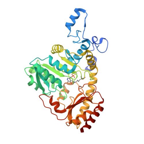 | |
UniProt | |||||
Find proteins for A0A0H3GVN6 (Klebsiella pneumoniae subsp. pneumoniae (strain HS11286)) Explore A0A0H3GVN6 Go to UniProtKB: A0A0H3GVN6 | |||||
Entity Groups | |||||
| Sequence Clusters | 30% Identity50% Identity70% Identity90% Identity95% Identity100% Identity | ||||
| UniProt Group | A0A0H3GVN6 | ||||
Sequence AnnotationsExpand | |||||
Reference Sequence | |||||
| Ligands 2 Unique | |||||
|---|---|---|---|---|---|
| ID | Chains | Name / Formula / InChI Key | 2D Diagram | 3D Interactions | |
| MPD Download:Ideal Coordinates CCD File | G [auth A], L [auth C], O [auth D] | (4S)-2-METHYL-2,4-PENTANEDIOL C6 H14 O2 SVTBMSDMJJWYQN-YFKPBYRVSA-N |  | ||
| CL Download:Ideal Coordinates CCD File | E [auth A] F [auth A] H [auth B] I [auth B] J [auth C] | CHLORIDE ION Cl VEXZGXHMUGYJMC-UHFFFAOYSA-M |  | ||
| Modified Residues 1 Unique | |||||
|---|---|---|---|---|---|
| ID | Chains | Type | Formula | 2D Diagram | Parent |
| LLP Query on LLP | A, B, C, D | L-PEPTIDE LINKING | C14 H22 N3 O7 P |  | LYS |
| Length ( Å ) | Angle ( ˚ ) |
|---|---|
| a = 82.61 | α = 90 |
| b = 131.14 | β = 99.93 |
| c = 85.44 | γ = 90 |
| Software Name | Purpose |
|---|---|
| PHENIX | refinement |
| XDS | data reduction |
| PDB_EXTRACT | data extraction |
| XSCALE | data scaling |
| MoRDa | phasing |
| Funding Organization | Location | Grant Number |
|---|---|---|
| National Institutes of Health/National Institute Of Allergy and Infectious Diseases (NIH/NIAID) | United States | -- |


