Structures of Fission Yeast Inositol Pyrophosphate Kinase Asp1 in Ligand-Free, Substrate-Bound, and Product-Bound States.
Benjamin, B., Goldgur, Y., Jork, N., Jessen, H.J., Schwer, B., Shuman, S.(2022) mBio 13: e0308722-e0308722
- PubMed: 36468882 Search on PubMedSearch on PubMed Central
- DOI: https://doi.org/10.1128/mbio.03087-22
- Primary Citation Related Structures:
8E1H, 8E1I, 8E1J, 8E1S, 8E1T, 8E1V - PubMed Abstract:
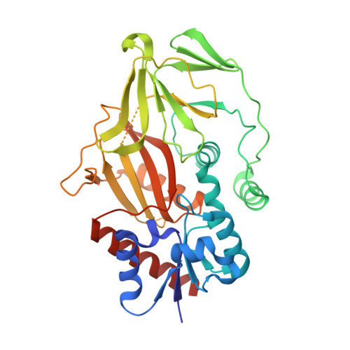
Expression of the fission yeast Schizosaccharomyces pombe phosphate regulon is sensitive to the intracellular level of the inositol pyrophosphate signaling molecule 1,5-IP 8 . IP 8 dynamics are determined by Asp1, a bifunctional enzyme consisting of an N-terminal kinase domain and a C-terminal pyrophosphatase domain that catalyze IP 8 synthesis and catabolism, respectively. Here, we report structures of the Asp1 kinase domain, crystallized with two protomers in the asymmetric unit, one of which was complexed with ligands (ADPNP, ADP, or ATP; Mg 2+ or Mn 2+ ; IP 6 , 5-IP 7 , or 1,5-IP 8 ) and the other which was ligand-free. The ligand-free enzyme adopts an "open" conformation that allows ingress of substrates and egress of products. ADPNP, ADP, and ATP and associated metal ions occupy a deep phospho-donor pocket in the active site. IP 6 or 5-IP 7 engagement above the nucleotide favors adoption of a "closed" conformation, in which surface protein segments undergo movement and a disordered-to-ordered transition to form an inositol polyphosphate-binding site. In a structure mimetic of the kinase Michaelis complex, the anionic 5-IP 7 phosphates are encaged by an ensemble of nine cationic amino acids: Lys43, Arg223, Lys224, Lys260, Arg274, Arg285, Lys290, Arg293, and Lys341. Alanine mutagenesis of amino acids that contact the adenosine nucleoside of the ATP donor underscored the contributions of Asp258 interaction with the ribose 3'-OH and of Glu248 with adenine- N 6 . Changing Glu248 to Gln elicited a gain of function whereby the kinase became adept at using GTP as phosphate donor. Wild-type Asp1 kinase can utilize N 6 -benzyl-ATP as phosphate donor. IMPORTANCE The inositol pyrophosphate signaling molecule 1,5-IP 8 modulates fission yeast phosphate homeostasis via its action as an agonist of RNA 3'-processing and transcription termination. Cellular IP 8 levels are determined by Asp1, a bifunctional enzyme composed of an N-terminal kinase and a C-terminal pyrophosphatase domain. Here, we present a series of crystal structures of the Asp1 kinase domain, in a ligand-free state and in complexes with nucleotides ADPNP, ADP, and ATP, divalent cations magnesium and manganese, and inositol polyphosphates IP 6 , 5-IP 7 , and 1,5-IP 8 . Substrate binding elicits a switch from open to closed conformations, entailing a disordered-to-ordered transition and a rearrangement or movement of two peptide segments that form a binding site for the phospho-acceptor. Our structures, along with structure-guided mutagenesis, fortify understanding of the mechanism and substrate specificity of Asp1 kinase, and they extend and complement structural and functional studies of the orthologous human kinase PPIP5K2.
- Molecular Biology Program, Memorial Sloan Kettering Cancer Centergrid.51462.34, New York, New York, USA.
Organizational Affiliation: