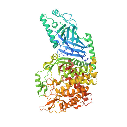
8EPR

Co-crystal structure of Chaetomium glucosidase with compound 19


Experimental Data Snapshot

  • Method: X-RAY DIFFRACTION
  • Resolution: 1.99 Å
  • R-Value Free: 0.225 
  • R-Value Work: 0.187 
  • R-Value Observed: 0.189 

Starting Model: experimental
View more details

wwPDB Validation   3D Report Full Report


Ligand Structure Quality Assessment 


This is version 1.3 of the entry. See complete history


Literature

Structure-Based Design of Potent Iminosugar Inhibitors of Endoplasmic Reticulum alpha-Glucosidase I with Anti-SARS-CoV-2 Activity.

Karade, S.S.Franco, E.J.Rojas, A.C.Hanrahan, K.C.Kolesnikov, A.Yu, W.MacKerell Jr., A.D.Hill, D.C.Weber, D.J.Brown, A.N.Treston, A.M.Mariuzza, R.A.

(2023) J Med Chem 66: 2744-2760

  • DOI: https://doi.org/10.1021/acs.jmedchem.2c01750

  • PubMed Abstract: 

    Enveloped viruses depend on the host endoplasmic reticulum (ER) quality control (QC) machinery for proper glycoprotein folding. The endoplasmic reticulum quality control (ERQC) enzyme α-glucosidase I (α-GluI) is an attractive target for developing broad-spectrum antivirals. We synthesized 28 inhibitors designed to interact with all four subsites of the α-GluI active site. These inhibitors are derivatives of the iminosugars 1-deoxynojirimycin (1-DNJ) and valiolamine. Crystal structures of ER α-GluI bound to 25 1-DNJ and three valiolamine derivatives revealed the basis for inhibitory potency. We established the structure-activity relationship (SAR) and used the Site Identification by Ligand Competitive Saturation (SILCS) method to develop a model for predicting α-GluI inhibition. We screened the compounds against SARS-CoV-2 in vitro to identify those with greater antiviral activity than the benchmark α-glucosidase inhibitor UV-4. These host-targeting compounds are candidates for investigation in animal models of SARS-CoV-2 and for testing against other viruses that rely on ERQC for correct glycoprotein folding.


  • Organizational Affiliation

    University of Maryland Institute for Bioscience and Biotechnology Research, Rockville, Maryland 20850, United States.


Macromolecules
Find similar proteins by:  (by identity cutoff)  |  3D Structure
Entity ID: 1
MoleculeChains Sequence LengthOrganismDetailsImage
Chaetomium alpha glucosidase
A, B
819Thermochaetoides thermophila DSM 1495Mutation(s): 0 
Gene Names: CTHT_0061620
EC: 3.2.1.106
UniProt
Find proteins for G0SFD1 (Chaetomium thermophilum (strain DSM 1495 / CBS 144.50 / IMI 039719))
Explore G0SFD1 
Go to UniProtKB:  G0SFD1
Entity Groups  
Sequence Clusters30% Identity50% Identity70% Identity90% Identity95% Identity100% Identity
UniProt GroupG0SFD1
Glycosylation
Glycosylation Sites: 1
Sequence Annotations
Expand
  • Reference Sequence
Small Molecules
Ligands 5 Unique
IDChains Name / Formula / InChI Key2D Diagram3D Interactions
WSW (Subject of Investigation/LOI)
Query on WSW

Download Ideal Coordinates CCD File 
C [auth A],
J [auth B]
(2R,3R,4R,5S)-2-(hydroxymethyl)-1-{[3-({[(5M)-3-(methanesulfonyl)-5-(pyridazin-3-yl)phenyl]amino}methyl)phenyl]methyl}piperidine-3,4,5-triol
C25 H30 N4 O6 S
ITFAXCBRKSPWOG-ZFFYZDHPSA-N
NAG
Query on NAG

Download Ideal Coordinates CCD File 
M [auth B]2-acetamido-2-deoxy-beta-D-glucopyranose
C8 H15 N O6
OVRNDRQMDRJTHS-FMDGEEDCSA-N
BTB
Query on BTB

Download Ideal Coordinates CCD File 
K [auth B]2-[BIS-(2-HYDROXY-ETHYL)-AMINO]-2-HYDROXYMETHYL-PROPANE-1,3-DIOL
C8 H19 N O5
OWMVSZAMULFTJU-UHFFFAOYSA-N
SO4
Query on SO4

Download Ideal Coordinates CCD File 
G [auth A]
H [auth A]
I [auth A]
O [auth B]
P [auth B]
G [auth A],
H [auth A],
I [auth A],
O [auth B],
P [auth B],
Q [auth B],
R [auth B]
SULFATE ION
O4 S
QAOWNCQODCNURD-UHFFFAOYSA-L
GOL
Query on GOL

Download Ideal Coordinates CCD File 
D [auth A],
E [auth A],
F [auth A],
L [auth B],
N [auth B]
GLYCEROL
C3 H8 O3
PEDCQBHIVMGVHV-UHFFFAOYSA-N
Experimental Data & Validation

Experimental Data

  • Method: X-RAY DIFFRACTION
  • Resolution: 1.99 Å
  • R-Value Free: 0.225 
  • R-Value Work: 0.187 
  • R-Value Observed: 0.189 
  • Space Group: I 2 2 2
Unit Cell:
Length ( Å )Angle ( ˚ )
a = 135.625α = 90
b = 178.74β = 90
c = 179.851γ = 90
Software Package:
Software NamePurpose
REFMACrefinement
PDB_EXTRACTdata extraction
HKL-2000data reduction
HKL-2000data scaling
PHASERphasing

Structure Validation

View Full Validation Report


Ligand Structure Quality Assessment 


Entry History & Funding Information

Deposition Data


Funding OrganizationLocationGrant Number
Not funded--

Revision History  (Full details and data files)

  • Version 1.0: 2023-02-22
    Type: Initial release
  • Version 1.1: 2023-03-01
    Changes: Database references
  • Version 1.2: 2023-10-25
    Changes: Data collection, Refinement description
  • Version 1.3: 2024-10-23
    Changes: Structure summary