


Crystal Structure of HIV-1 LM/HT CLADE A/E CRF01 GP120 Core in Complex with TFH-II-151
Tolbert, W.D., Nguyen, D.N., Pazgier, M.To be published.
Experimental Data Snapshot
Starting Model: experimental
View more details
Entity ID: 1 | |||||
|---|---|---|---|---|---|
| Molecule | Chains | Sequence Length | Organism | Details | Image |
| HIV-1 LM/HT Clade A/E CRF01 gp120 core | 355 | Human immunodeficiency virus 1 | Mutation(s): 7 Gene Names: HIV-1 Env | 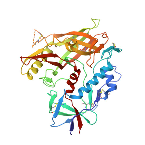 | |
UniProt | |||||
Find proteins for A0A0M3KKW9 (Human immunodeficiency virus type 1) Explore A0A0M3KKW9 Go to UniProtKB: A0A0M3KKW9 | |||||
Entity Groups | |||||
| Sequence Clusters | 30% Identity50% Identity70% Identity90% Identity95% Identity100% Identity | ||||
| UniProt Group | A0A0M3KKW9 | ||||
Glycosylation | |||||
| Glycosylation Sites: 13 | |||||
Sequence AnnotationsExpand | |||||
Reference Sequence | |||||
| Ligands 4 Unique | |||||
|---|---|---|---|---|---|
| ID | Chains | Name / Formula / InChI Key | 2D Diagram | 3D Interactions | |
| Z7N (Subject of Investigation/LOI) Download:Ideal Coordinates CCD File | O [auth A] | (3S,5R)-N-(4-chloro-3-fluorophenyl)-5-(hydroxymethyl)-1-[(3R,5S)-3,4,5-trimethylpiperazine-1-carbonyl]piperidine-3-carboxamide C21 H30 Cl F N4 O3 ICLRAUCRFDHWBU-QXSJWSMHSA-N |  | ||
| EPE Download:Ideal Coordinates CCD File | P [auth A] | 4-(2-HYDROXYETHYL)-1-PIPERAZINE ETHANESULFONIC ACID C8 H18 N2 O4 S JKMHFZQWWAIEOD-UHFFFAOYSA-N |  | ||
| NAG Download:Ideal Coordinates CCD File | B [auth A] C [auth A] D [auth A] E [auth A] F [auth A] | 2-acetamido-2-deoxy-beta-D-glucopyranose C8 H15 N O6 OVRNDRQMDRJTHS-FMDGEEDCSA-N |  | ||
| CL Download:Ideal Coordinates CCD File | Q [auth A], R [auth A], S [auth A] | CHLORIDE ION Cl VEXZGXHMUGYJMC-UHFFFAOYSA-M |  | ||
| Length ( Å ) | Angle ( ˚ ) |
|---|---|
| a = 61.418 | α = 90 |
| b = 65.997 | β = 90 |
| c = 90.826 | γ = 90 |
| Software Name | Purpose |
|---|---|
| PHENIX | refinement |
| HKL-3000 | data reduction |
| HKL-3000 | data scaling |
| PHASER | phasing |
| Funding Organization | Location | Grant Number |
|---|---|---|
| National Institutes of Health/National Institute Of Allergy and Infectious Diseases (NIH/NIAID) | United States | R01AI129769 |


