8GOS | pdb_00008gos

Crystal structure of fluorescent protein RasM


Experimental Data Snapshot

  • Method: X-RAY DIFFRACTION
  • Resolution: 1.01 Å
  • R-Value Free: 
    0.158 (Depositor), 0.159 (DCC) 
  • R-Value Work: 
    0.138 (Depositor), 0.139 (DCC) 
  • R-Value Observed: 
    0.139 (Depositor) 

wwPDB Validation 3D Report Full Report

Validation slider image for 8GOS

This is version 2.0 of the entry. See complete history

Literature

Beat-frequency-resolved two-dimensional electronic spectroscopy: disentangling vibrational coherences in artificial fluorescent proteins with sub-10-fs visible laser pulses.

Tsubouchi, M.Ishii, N.Kagotani, Y.Shimizu, R.Fujita, T.Adachi, M.Itakura, R.

(2023) Opt Express 31: 6890-6906

  • DOI: https://doi.org/10.1364/OE.480505
  • Primary Citation Related Structures: 
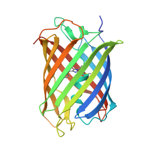
    8GOS

  • PubMed Abstract: 

    We perform a beat-frequency-resolved analysis for two-dimensional electronic spectroscopy using a high-speed and stable 2D electronic spectrometer and few-cycle visible laser pulses to disentangle the vibrational coherences in an artificial fluorescent protein. We develop a highly stable ultrashort light source that generates 5.3-fs visible pulses with a pulse energy of 4.7 µJ at a repetition rate of 10 kHz using multi-plate pulse compression and laser filamentation in a gas cell. The above-5.3-fs laser pulses together with a high-speed multichannel detector enable us to measure a series of 2D electronic spectra, which are resolved in terms of beat frequency related to vibrational coherence. We successfully extract the discrete vibrational peaks behind the inhomogeneous broadening in the absorption spectra and the vibrational quantum beats of the excited electronic state behind the strong incoherent population background in the typical 2D electronic spectra.

Macromolecule Content 

  • Total Structure Weight: 51.63 kDa 
  • Atom Count: 4,129 
  • Modeled Residue Count: 430 
  • Deposited Residue Count: 452 
  • Unique protein chains: 1

Macromolecules

Find similar proteins by:|  3D Structure
Entity ID: 1
MoleculeChains  Sequence LengthOrganismDetailsImage
RasM
A, B
226Discosoma sp.Mutation(s): 0 
UniProt
Find proteins for D0VWW2 (Discosoma sp.)
Explore D0VWW2 
Go to UniProtKB:  D0VWW2
Entity Groups
Sequence Clusters30% Identity50% Identity70% Identity90% Identity95% Identity100% Identity
UniProt GroupD0VWW2
Sequence Annotations
Expand
Reference Sequence

Small Molecules

Modified Residues  1 Unique
IDChains TypeFormula2D DiagramParent
NRQ
Query on NRQ
A, B
L-PEPTIDE LINKINGC16 H17 N3 O4 SMET, TYR, GLY

Experimental Data & Validation

Experimental Data

  • Method: X-RAY DIFFRACTION
  • Resolution: 1.01 Å
  • R-Value Free:  0.158 (Depositor), 0.159 (DCC) 
  • R-Value Work:  0.138 (Depositor), 0.139 (DCC) 
  • R-Value Observed: 0.139 (Depositor) 
Space Group: P 21 21 21
Unit Cell:
Length ( Å )Angle ( ˚ )
a = 59.394α = 90
b = 62.328β = 90
c = 107.819γ = 90
Software Package:
Software NamePurpose
PHENIXrefinement
XDSdata reduction
pointlessdata scaling
PHENIXphasing

Structure Validation

View Full Validation Report


Entry History 

& Funding Information

Deposition Data


Funding OrganizationLocationGrant Number
Ministry of Education, Culture, Sports, Science and Technology (Japan)JapanJPMXS0120330644

Revision History  (Full details and data files)

  • Version 1.0: 2023-09-27
    Type: Initial release
  • Version 1.1: 2023-11-15
    Changes: Data collection
  • Version 2.0: 2026-03-18
    Changes: Polymer sequence, Structure summary