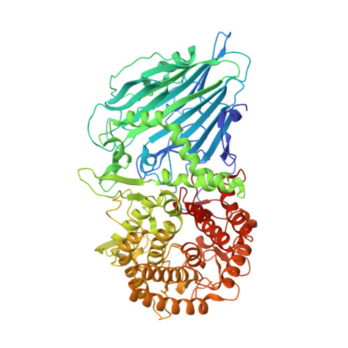
8I5U

Crystal structure of TxGH116 D593N acid/base mutant from Thermoanaerobacterium xylanolyticum with laminaribiose


Experimental Data Snapshot

  • Method: X-RAY DIFFRACTION
  • Resolution: 1.40 Å
  • R-Value Free: 0.171 
  • R-Value Work: 0.153 
  • R-Value Observed: 0.154 

Starting Model: experimental
View more details

wwPDB Validation   3D Report Full Report


This is version 1.2 of the entry. See complete history


Literature

Reaction Mechanism of Glycoside Hydrolase Family 116 Utilizes Perpendicular Protonation.

Pengthaisong, S.Piniello, B.Davies, G.J.Rovira, C.Ketudat Cairns, J.R.

(2023) ACS Catal 13: 5850-5863

  • DOI: https://doi.org/10.1021/acscatal.3c00620
  • Primary Citation of Related Structures:  
    8I5O, 8I5P, 8I5Q, 8I5R, 8I5S, 8I5T, 8I5U

  • PubMed Abstract: 

    Retaining glycoside hydrolases use acid/base catalysis with an enzymatic acid/base protonating the glycosidic bond oxygen to facilitate leaving-group departure alongside attack by a catalytic nucleophile to form a covalent intermediate. Generally, this acid/base protonates the oxygen laterally with respect to the sugar ring, which places the catalytic acid/base and nucleophile carboxylates within about 4.5-6.5 Å of each other. However, in glycoside hydrolase (GH) family 116, including disease-related human acid β-glucosidase 2 (GBA2), the distance between the catalytic acid/base and the nucleophile is around 8 Å (PDB: 5BVU) and the catalytic acid/base appears to be above the plane of the pyranose ring, rather than being lateral to that plane, which could have catalytic consequences. However, no structure of an enzyme-substrate complex is available for this GH family. Here, we report the structures of Thermoanaerobacterium xylanolyticum β-glucosidase ( Tx GH116) D593N acid/base mutant in complexes with cellobiose and laminaribiose and its catalytic mechanism. We confirm that the amide hydrogen bonding to the glycosidic oxygen is in a perpendicular rather than lateral orientation. Quantum mechanics/molecular mechanics (QM/MM) simulations of the glycosylation half-reaction in wild-type Tx GH116 indicate that the substrate binds with the nonreducing glucose residue in an unusual relaxed 4 C 1 chair at the -1 subsite. Nevertheless, the reaction can still proceed through a 4 H 3 half-chair transition state, as in classical retaining β-glucosidases, as the catalytic acid D593 protonates the perpendicular electron pair. The glucose C6OH is locked in a gauche , trans orientation with respect to the C5-O5 and C4-C5 bonds to facilitate perpendicular protonation. These data imply a unique protonation trajectory in Clan-O glycoside hydrolases, which has strong implications for the design of inhibitors specific to either lateral protonators, such as human GBA1, or perpendicular protonators, such as human GBA2.


  • Organizational Affiliation

    School of Chemistry, Institute of Science, Suranaree University of Technology, Nakhon Ratchasima 30000, Thailand.


Macromolecules
Find similar proteins by:  (by identity cutoff)  |  3D Structure
Entity ID: 1
MoleculeChains Sequence LengthOrganismDetailsImage
beta-glucosidase799Thermoanaerobacterium xylanolyticum LX-11Mutation(s): 1 
Gene Names: Thexy_2211
EC: 3.2.1.21 (PDB Primary Data), 3.2.1.45 (UniProt)
UniProt
Find proteins for F6BL85 (Thermoanaerobacterium xylanolyticum (strain ATCC 49914 / DSM 7097 / LX-11))
Explore F6BL85 
Go to UniProtKB:  F6BL85
Entity Groups  
Sequence Clusters30% Identity50% Identity70% Identity90% Identity95% Identity100% Identity
UniProt GroupF6BL85
Sequence Annotations
Expand
  • Reference Sequence
Oligosaccharides

Help

Entity ID: 2
MoleculeChains Length2D Diagram Glycosylation3D Interactions
beta-D-glucopyranose-(1-3)-beta-D-glucopyranose
B
2N/A
Glycosylation Resources
GlyTouCan:  G36535HU
GlyCosmos:  G36535HU
Small Molecules
Ligands 3 Unique
IDChains Name / Formula / InChI Key2D Diagram3D Interactions
GOL
Query on GOL

Download Ideal Coordinates CCD File 
C [auth A]
D [auth A]
E [auth A]
F [auth A]
G [auth A]
C [auth A],
D [auth A],
E [auth A],
F [auth A],
G [auth A],
H [auth A],
I [auth A],
J [auth A],
K [auth A],
L [auth A],
M [auth A],
N [auth A],
O [auth A],
P [auth A],
Q [auth A],
R [auth A]
GLYCEROL
C3 H8 O3
PEDCQBHIVMGVHV-UHFFFAOYSA-N
EDO
Query on EDO

Download Ideal Coordinates CCD File 
T [auth A],
U [auth A]
1,2-ETHANEDIOL
C2 H6 O2
LYCAIKOWRPUZTN-UHFFFAOYSA-N
CA
Query on CA

Download Ideal Coordinates CCD File 
S [auth A]CALCIUM ION
Ca
BHPQYMZQTOCNFJ-UHFFFAOYSA-N
Biologically Interesting Molecules (External Reference) 1 Unique
Experimental Data & Validation

Experimental Data

  • Method: X-RAY DIFFRACTION
  • Resolution: 1.40 Å
  • R-Value Free: 0.171 
  • R-Value Work: 0.153 
  • R-Value Observed: 0.154 
  • Space Group: P 21 21 2
Unit Cell:
Length ( Å )Angle ( ˚ )
a = 177.765α = 90
b = 54.129β = 90
c = 83.33γ = 90
Software Package:
Software NamePurpose
REFMACrefinement
HKL-2000data collection
HKL-2000data scaling
MOLREPphasing
Cootmodel building
PDB_EXTRACTdata extraction
HKL-2000data reduction

Structure Validation

View Full Validation Report


Entry History & Funding Information

Deposition Data


Funding OrganizationLocationGrant Number
Other governmentThailandThailand Research Fund
Other governmentThailandSuranaree University of Technology and Office of the Higher Education Commission under NRU Project of Thailand
Other governmentThailandThailand Science Research and Innovation

Revision History  (Full details and data files)

  • Version 1.0: 2023-05-03
    Type: Initial release
  • Version 1.1: 2023-05-31
    Changes: Database references
  • Version 1.2: 2023-11-29
    Changes: Data collection, Refinement description