


Structure of colicin Ib Channel domain at 3.0 Angstroms resolution
Yang, J.To be published.
Experimental Data Snapshot
Starting Model: experimental
View more details
wwPDB Validation 3D Report Full Report

Entity ID: 1 | |||||
|---|---|---|---|---|---|
| Molecule | Chains | Sequence Length | Organism | Details | Image |
| Colicin 1B | 205 | Shigella flexneri | Mutation(s): 0 Gene Names: CBL31_01480 | 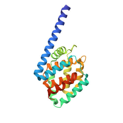 | |
| Length ( Å ) | Angle ( ˚ ) |
|---|---|
| a = 91.994 | α = 90 |
| b = 91.994 | β = 90 |
| c = 113.896 | γ = 90 |
| Software Name | Purpose |
|---|---|
| PHENIX | refinement |
| HKL-2000 | data collection |
| HKL-2000 | data scaling |
| PHENIX | model building |
| Funding Organization | Location | Grant Number |
|---|---|---|
| National Science Council (NSC, Taiwan) | Taiwan | 110-2311-B-005-005-MY3 |


