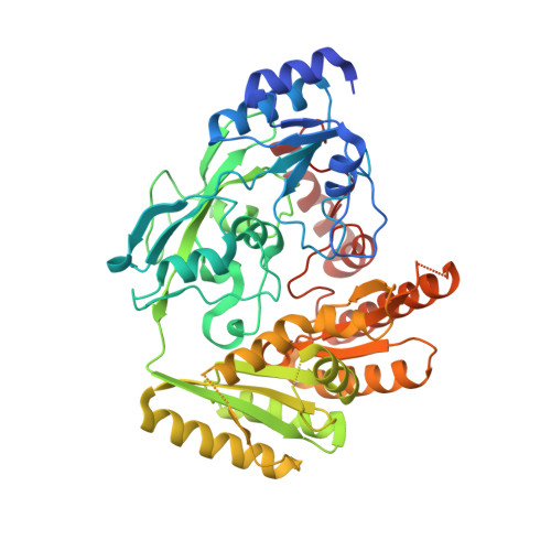
8JDZ

Crystal structure of mLDHD in complex with 2-keto-3-methylvaleric acid


Experimental Data Snapshot

  • Method: X-RAY DIFFRACTION
  • Resolution: 1.56 Å
  • R-Value Free: 0.205 
  • R-Value Work: 0.180 
  • R-Value Observed: 0.182 

Starting Model: experimental
View more details

wwPDB Validation   3D Report Full Report


Ligand Structure Quality Assessment 


This is version 1.1 of the entry. See complete history


Literature

Lactate dehydrogenase D is a general dehydrogenase for D-2-hydroxyacids and is associated with D-lactic acidosis.

Jin, S.Chen, X.Yang, J.Ding, J.

(2023) Nat Commun 14: 6638-6638

  • DOI: https://doi.org/10.1038/s41467-023-42456-3
  • Primary Citation of Related Structures:  
    8JDB, 8JDC, 8JDD, 8JDE, 8JDF, 8JDG, 8JDN, 8JDO, 8JDP, 8JDQ, 8JDR, 8JDS, 8JDT, 8JDU, 8JDV, 8JDX, 8JDY, 8JDZ

  • PubMed Abstract: 

    Mammalian lactate dehydrogenase D (LDHD) catalyzes the oxidation of D-lactate to pyruvate. LDHD mutations identified in patients with D-lactic acidosis lead to deficient LDHD activity. Here, we perform a systematic biochemical study of mouse LDHD (mLDHD) and determine the crystal structures of mLDHD in FAD-bound form and in complexes with FAD, Mn 2+ and a series of substrates or products. We demonstrate that mLDHD is an Mn 2+ -dependent general dehydrogenase which exhibits catalytic activity for D-lactate and other D-2-hydroxyacids containing hydrophobic moieties, but no activity for their L-isomers or D-2-hydroxyacids containing hydrophilic moieties. The substrate-binding site contains a positively charged pocket to bind the common glycolate moiety and a hydrophobic pocket with some elasticity to bind the varied hydrophobic moieties of substrates. The structural and biochemical data together reveal the molecular basis for the substrate specificity and catalytic mechanism of LDHD, and the functional roles of mutations in the pathogenesis of D-lactic acidosis.


  • Organizational Affiliation

    State Key Laboratory of Molecular Biology, Shanghai Institute of Biochemistry and Cell Biology, Center for Excellence in Molecular Cell Science, University of Chinese Academy of Sciences, Chinese Academy of Sciences, 320 Yue-Yang Road, Shanghai, 200031, China.


Macromolecules
Find similar proteins by:  (by identity cutoff)  |  3D Structure
Entity ID: 1
MoleculeChains Sequence LengthOrganismDetailsImage
Probable D-lactate dehydrogenase, mitochondrial471Mus musculusMutation(s): 0 
Gene Names: Ldhd
EC: 1.1.2.4
UniProt
Find proteins for Q7TNG8 (Mus musculus)
Explore Q7TNG8 
Go to UniProtKB:  Q7TNG8
Entity Groups  
Sequence Clusters30% Identity50% Identity70% Identity90% Identity95% Identity100% Identity
UniProt GroupQ7TNG8
Sequence Annotations
Expand
  • Reference Sequence
Experimental Data & Validation

Experimental Data

  • Method: X-RAY DIFFRACTION
  • Resolution: 1.56 Å
  • R-Value Free: 0.205 
  • R-Value Work: 0.180 
  • R-Value Observed: 0.182 
  • Space Group: I 2 2 2
Unit Cell:
Length ( Å )Angle ( ˚ )
a = 82.07α = 90
b = 102.58β = 90
c = 122.94γ = 90
Software Package:
Software NamePurpose
PHENIXrefinement
Aimlessdata scaling
xia2data reduction
PHENIXphasing

Structure Validation

View Full Validation Report


Ligand Structure Quality Assessment 


Entry History & Funding Information

Deposition Data


Funding OrganizationLocationGrant Number
National Natural Science Foundation of China (NSFC)China31870723
Chinese Academy of SciencesChinaXDB37030305

Revision History  (Full details and data files)

  • Version 1.0: 2023-10-18
    Type: Initial release
  • Version 1.1: 2024-02-21
    Changes: Database references