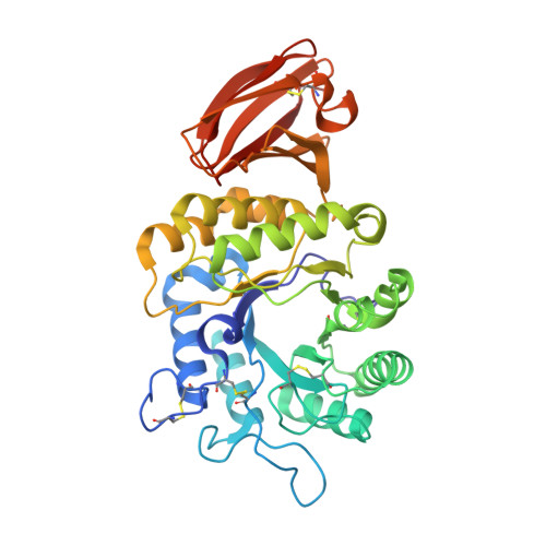
8K7E

Crystal structure of human lysosomal alpha-galactosidase A in complex with (2R,3R,4S,5R)-2-(aminomethyl)-5-(hydroxymethyl)pyrrolidine-3,4-diol


Experimental Data Snapshot

  • Method: X-RAY DIFFRACTION
  • Resolution: 2.20 Å
  • R-Value Free: 0.192 
  • R-Value Work: 0.160 
  • R-Value Observed: 0.161 

Starting Model: experimental
View more details

wwPDB Validation   3D Report Full Report


Ligand Structure Quality Assessment 


This is version 1.0 of the entry. See complete history


Literature

Mechanistic Insights into Dibasic Iminosugars as pH-Selective Pharmacological Chaperones to Stabilize Human alpha-Galactosidase.

Li, H.Y.Lin, H.Y.Chang, S.K.Chiu, Y.T.Hou, C.C.Ko, T.P.Huang, K.F.Niu, D.M.Cheng, W.C.

(2024) JACS Au 4: 908-918

  • DOI: https://doi.org/10.1021/jacsau.3c00684
  • Primary Citation of Related Structures:  
    8K7D, 8K7E, 8K7F, 8K7G, 8K7H, 8K7I, 8K7J, 8K7K, 8K7L

  • PubMed Abstract: 

    The use of pharmacological chaperones (PCs) to stabilize specific enzymes and impart a therapeutic benefit is an emerging strategy in drug discovery. However, designing molecules that can bind optimally to their targets at physiological pH remains a major challenge. Our previous study found that dibasic polyhydroxylated pyrrolidine 5 exhibited superior pH-selective inhibitory activity and chaperoning activity for human α-galactosidase A (α-Gal A) compared with its monobasic parent molecule, 4 . To further investigate the role of different C-2 moieties on the pH-selectivity and protecting effects of these compounds, we designed and synthesized a library of monobasic and dibasic iminosugars, screened them for α-Gal A-stabilizing activity using thermal shift and heat-induced denaturation assays, and characterized the mechanistic basis for this stabilization using X-ray crystallography and binding assays. We noted that the dibasic iminosugars 5 and 20 protect α-Gal A from denaturation and inactivation at lower concentrations than monobasic or other N -substituted derivatives; a finding attributed to the nitrogen on the C-2 methylene of 5 and 20 , which forms the bifurcated salt bridges (BSBs) with two carboxyl residues, E203 and D231. Additionally, the formation of BSBs at pH 7.0 and the electrostatic repulsion between the vicinal ammonium cations of dibasic iminosugars at pH 4.5 are responsible for their pH-selective binding to α-Gal A. Moreover, compounds 5 and 20 demonstrated promising results in improving enzyme replacement therapy and exhibited significant chaperoning effects in Fabry cells. These findings suggest amino-iminosugars 5 and 20 as useful models to demonstrate how an additional exocyclic amino group can improve their pH-selectivity and protecting effects, providing new insights for the design of pH-selective PCs.


  • Organizational Affiliation

    Genomics Research Center, Academia Sinica, 128, Section 2, Academia Road, Nankang, Taipei 115201, Taiwan.


Macromolecules
Find similar proteins by:  (by identity cutoff)  |  3D Structure
Entity ID: 1
MoleculeChains Sequence LengthOrganismDetailsImage
Alpha-galactosidase A
A, B
398Homo sapiensMutation(s): 0 
Gene Names: GLA
EC: 3.2.1.22
UniProt & NIH Common Fund Data Resources
Find proteins for P06280 (Homo sapiens)
Explore P06280 
Go to UniProtKB:  P06280
PHAROS:  P06280
GTEx:  ENSG00000102393 
Entity Groups  
Sequence Clusters30% Identity50% Identity70% Identity90% Identity95% Identity100% Identity
UniProt GroupP06280
Glycosylation
Glycosylation Sites: 2Go to GlyGen: P06280-1
Sequence Annotations
Expand
  • Reference Sequence
Oligosaccharides

Help

Entity ID: 2
MoleculeChains Length2D Diagram Glycosylation3D Interactions
2-acetamido-2-deoxy-beta-D-glucopyranose-(1-4)-2-acetamido-2-deoxy-beta-D-glucopyranose
C, D
2N-Glycosylation
Glycosylation Resources
GlyTouCan:  G42666HT
GlyCosmos:  G42666HT
GlyGen:  G42666HT
Entity ID: 3
MoleculeChains Length2D Diagram Glycosylation3D Interactions
alpha-D-mannopyranose-(1-3)-beta-D-mannopyranose-(1-4)-2-acetamido-2-deoxy-beta-D-glucopyranose-(1-4)-2-acetamido-2-deoxy-beta-D-glucopyranose
E
4N-Glycosylation
Glycosylation Resources
GlyTouCan:  G81315DD
GlyCosmos:  G81315DD
GlyGen:  G81315DD
Small Molecules
Ligands 3 Unique
IDChains Name / Formula / InChI Key2D Diagram3D Interactions
NAG
Query on NAG

Download Ideal Coordinates CCD File 
F [auth A],
I [auth B]
2-acetamido-2-deoxy-beta-D-glucopyranose
C8 H15 N O6
OVRNDRQMDRJTHS-FMDGEEDCSA-N
VLN (Subject of Investigation/LOI)
Query on VLN

Download Ideal Coordinates CCD File 
G [auth A],
J [auth B]
(2~{R},3~{R},4~{S},5~{R})-2-(aminomethyl)-5-(hydroxymethyl)pyrrolidine-3,4-diol
C6 H14 N2 O3
NJOZFAFEQVQFJI-KAZBKCHUSA-N
SO4
Query on SO4

Download Ideal Coordinates CCD File 
H [auth A],
K [auth B]
SULFATE ION
O4 S
QAOWNCQODCNURD-UHFFFAOYSA-L
Experimental Data & Validation

Experimental Data

  • Method: X-RAY DIFFRACTION
  • Resolution: 2.20 Å
  • R-Value Free: 0.192 
  • R-Value Work: 0.160 
  • R-Value Observed: 0.161 
  • Space Group: P 32 2 1
Unit Cell:
Length ( Å )Angle ( ˚ )
a = 90.37α = 90
b = 90.37β = 90
c = 216.423γ = 120
Software Package:
Software NamePurpose
PHENIXrefinement
HKL-2000data reduction
HKL-2000data scaling
MOLREPphasing

Structure Validation

View Full Validation Report


Ligand Structure Quality Assessment 


Entry History & Funding Information

Deposition Data


Funding OrganizationLocationGrant Number
Ministry of Science and Technology (MoST, Taiwan)Taiwan--
Academia Sinica (Taiwan)Taiwan--

Revision History  (Full details and data files)

  • Version 1.0: 2024-04-10
    Type: Initial release