8PZK | pdb_00008pzk

Crystal structure of the Orange Carotenoid Protein 2 (OCP2) from Gloeocapsa sp. PCC 7428


Experimental Data Snapshot

  • Method: X-RAY DIFFRACTION
  • Resolution: 1.80 Å
  • R-Value Free: 
    0.248 (Depositor), 0.249 (DCC) 
  • R-Value Work: 
    0.197 (Depositor), 0.198 (DCC) 

Starting Model: experimental
View more details

wwPDB Validation 3D Report Full Report

Validation slider image for 8PZK

Ligand Structure Quality Assessment 


This is version 1.1 of the entry. See complete history

Literature

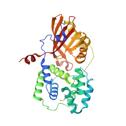
Structural framework for the understanding spectroscopic and functional signatures of the cyanobacterial Orange Carotenoid Protein families.

Sluchanko, N.N.Maksimov, E.G.Slonimskiy, Y.B.Varfolomeeva, L.A.Bukhanko, A.Y.Egorkin, N.A.Tsoraev, G.V.Khrenova, M.G.Ge, B.Qin, S.Boyko, K.M.Popov, V.O.

(2024) Int J Biol Macromol 254: 127874-127874

  • DOI: https://doi.org/10.1016/j.ijbiomac.2023.127874
  • Primary Citation Related Structures: 
    8PYH, 8PZK

  • PubMed Abstract: 

    The Orange Carotenoid Protein (OCP) is a unique photoreceptor crucial for cyanobacterial photoprotection. Best studied Synechocystis sp. PCC 6803 OCP belongs to the large OCP1 family. Downregulated by the Fluorescence Recovery Protein (FRP) in low-light, high-light-activated OCP1 binds to the phycobilisomes and performs non-photochemical quenching. Recently discovered families OCP2 and OCP3 remain structurally and functionally underexplored, and no systematic comparative studies have ever been conducted. Here we present two first crystal structures of OCP2 from morphoecophysiologically different cyanobacteria and provide their comprehensive structural, spectroscopic and functional comparison with OCP1, the recently described OCP3 and all-OCP ancestor. Structures enable correlation of spectroscopic signatures with the effective number of hydrogen and discovered here chalcogen bonds anchoring the ketocarotenoid in OCP, as well as with the rotation of the echinenone's β-ionone ring in the CTD. Structural data also helped rationalize the observed differences in OCP/FRP and OCP/phycobilisome functional interactions. These data are expected to foster OCP research and applications in optogenetics, targeted carotenoid delivery and cyanobacterial biomass engineering.


  • Organizational Affiliation
    • A.N. Bach Institute of Biochemistry, Federal Research Centre of Biotechnology of the Russian Academy of Sciences, Moscow 119071, Russia. Electronic address: nikolai.sluchanko@mail.ru.

Macromolecule Content 

  • Total Structure Weight: 36.39 kDa 
  • Atom Count: 2,541 
  • Modeled Residue Count: 300 
  • Deposited Residue Count: 321 
  • Unique protein chains: 1

Macromolecules

Find similar proteins by:|  3D Structure
Entity ID: 1
MoleculeChains  Sequence LengthOrganismDetailsImage
Orange carotenoid-binding protein321Gloeocapsa sp. PCC 7428Mutation(s): 1 
Gene Names: Glo7428_2455
UniProt
Find proteins for A0ACD6B8M9 (Gloeocapsa sp. PCC 7428)
Explore A0ACD6B8M9 
Go to UniProtKB:  A0ACD6B8M9
Entity Groups
Sequence Clusters30% Identity50% Identity70% Identity90% Identity95% Identity100% Identity
UniProt GroupA0ACD6B8M9
Sequence Annotations
Expand
Reference Sequence

Small Molecules

Ligands 4 Unique
IDChains Name / Formula / InChI Key2D Diagram3D Interactions
ECH
(Subject of Investigation/LOI)

Query on ECH


Download:Ideal Coordinates CCD File
B [auth A]beta,beta-caroten-4-one
C40 H54 O
QXNWZXMBUKUYMD-QQGJMDNJSA-N
TRS

Query on TRS


Download:Ideal Coordinates CCD File
C [auth A]2-AMINO-2-HYDROXYMETHYL-PROPANE-1,3-DIOL
C4 H12 N O3
LENZDBCJOHFCAS-UHFFFAOYSA-O
GOL

Query on GOL


Download:Ideal Coordinates CCD File
H [auth A]GLYCEROL
C3 H8 O3
PEDCQBHIVMGVHV-UHFFFAOYSA-N
AZI

Query on AZI


Download:Ideal Coordinates CCD File
D [auth A],
E [auth A],
F [auth A],
G [auth A],
I [auth A]
AZIDE ION
N3
IVRMZWNICZWHMI-UHFFFAOYSA-N
Modified Residues  1 Unique
IDChains TypeFormula2D DiagramParent
CSO
Query on CSO
A
L-PEPTIDE LINKINGC3 H7 N O3 SCYS

Experimental Data & Validation

Experimental Data

  • Method: X-RAY DIFFRACTION
  • Resolution: 1.80 Å
  • R-Value Free:  0.248 (Depositor), 0.249 (DCC) 
  • R-Value Work:  0.197 (Depositor), 0.198 (DCC) 
Space Group: P 43 21 2
Unit Cell:
Length ( Å )Angle ( ˚ )
a = 52.661α = 90
b = 52.661β = 90
c = 216.831γ = 90
Software Package:
Software NamePurpose
REFMACrefinement
Aimlessdata scaling
XDSdata reduction
PHASERphasing

Structure Validation

View Full Validation Report


Ligand Structure Quality Assessment 


Entry History 

& Funding Information

Deposition Data


Funding OrganizationLocationGrant Number
Ministry of Science and Higher Education of the Russian FederationRussian Federation075-15-2021-1354
Russian Science FoundationRussian Federation21-44-00005

Revision History  (Full details and data files)

  • Version 1.0: 2024-01-03
    Type: Initial release
  • Version 1.1: 2024-10-09
    Changes: Structure summary