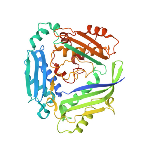
8QE0

Crystal structure of human MAT2a bound to S-Adenosylmethionine and Compound 12


Experimental Data Snapshot

  • Method: X-RAY DIFFRACTION
  • Resolution: 1.12 Å
  • R-Value Free: 0.187 
  • R-Value Work: 0.170 
  • R-Value Observed: 0.171 

wwPDB Validation   3D Report Full Report


Ligand Structure Quality Assessment 


This is version 1.1 of the entry. See complete history


Literature

Development of a Series of Pyrrolopyridone MAT2A Inhibitors.

Atkinson, S.J.Bagal, S.K.Argyrou, A.Askin, S.Cheung, T.Chiarparin, E.Coen, M.Collie, I.T.Dale, I.L.De Fusco, C.Dillman, K.Evans, L.Feron, L.J.Foster, A.J.Grondine, M.Kantae, V.Lamont, G.M.Lamont, S.Lynch, J.T.Nilsson Lill, S.Robb, G.R.Saeh, J.Schimpl, M.Scott, J.S.Smith, J.Srinivasan, B.Tentarelli, S.Vazquez-Chantada, M.Wagner, D.Walsh, J.J.Watson, D.Williamson, B.

(2024) J Med Chem 67: 4541-4559

  • DOI: https://doi.org/10.1021/acs.jmedchem.3c01860
  • Primary Citation of Related Structures:  
    8QDY, 8QDZ, 8QE0, 8QE1, 8QE2, 8QE3

  • PubMed Abstract: 

    The optimization of an allosteric fragment, discovered by differential scanning fluorimetry, to an in vivo MAT2a tool inhibitor is discussed. The structure-based drug discovery approach, aided by relative binding free energy calculations, resulted in AZ'9567 ( 21 ), a potent inhibitor in vitro with excellent preclinical pharmacokinetic properties. This tool showed a selective antiproliferative effect on methylthioadenosine phosphorylase (MTAP) KO cells, both in vitro and in vivo, providing further evidence to support the utility of MAT2a inhibitors as potential anticancer therapies for MTAP-deficient tumors.


  • Organizational Affiliation

    Oncology R&D, AstraZeneca, The Discovery Centre, Cambridge Biomedical Campus, 1 Francis Crick Avenue, Cambridge CB2 0AA, U.K.


Macromolecules
Find similar proteins by:  (by identity cutoff)  |  3D Structure
Entity ID: 1
MoleculeChains Sequence LengthOrganismDetailsImage
S-adenosylmethionine synthase isoform type-2397Homo sapiensMutation(s): 0 
Gene Names: MAT2AAMS2MATA2
EC: 2.5.1.6
UniProt & NIH Common Fund Data Resources
Find proteins for P31153 (Homo sapiens)
Explore P31153 
Go to UniProtKB:  P31153
PHAROS:  P31153
GTEx:  ENSG00000168906 
Entity Groups  
Sequence Clusters30% Identity50% Identity70% Identity90% Identity95% Identity100% Identity
UniProt GroupP31153
Sequence Annotations
Expand
  • Reference Sequence
Small Molecules
Ligands 2 Unique
IDChains Name / Formula / InChI Key2D Diagram3D Interactions
SAM
Query on SAM

Download Ideal Coordinates CCD File 
C [auth A]S-ADENOSYLMETHIONINE
C15 H22 N6 O5 S
MEFKEPWMEQBLKI-FCKMPRQPSA-N
U8R (Subject of Investigation/LOI)
Query on U8R

Download Ideal Coordinates CCD File 
B [auth A]4-[4-[bis(fluoranyl)methoxy]phenyl]-3-cyclopropyl-2~{H}-pyrazolo[4,3-b]pyridin-5-one
C16 H13 F2 N3 O2
DBMHKPKFMPEIGT-UHFFFAOYSA-N
Experimental Data & Validation

Experimental Data

  • Method: X-RAY DIFFRACTION
  • Resolution: 1.12 Å
  • R-Value Free: 0.187 
  • R-Value Work: 0.170 
  • R-Value Observed: 0.171 
  • Space Group: I 2 2 2
Unit Cell:
Length ( Å )Angle ( ˚ )
a = 68.179α = 90
b = 93.984β = 90
c = 116.409γ = 90
Software Package:
Software NamePurpose
BUSTERrefinement
STARANISOdata scaling
XDSdata reduction
AMoREphasing

Structure Validation

View Full Validation Report


Ligand Structure Quality Assessment 


Entry History & Funding Information

Deposition Data

  • Released Date: 2024-03-20 
  • Deposition Author(s): Schimpl, M.

Funding OrganizationLocationGrant Number
Not funded--

Revision History  (Full details and data files)

  • Version 1.0: 2024-03-20
    Type: Initial release
  • Version 1.1: 2024-04-10
    Changes: Database references