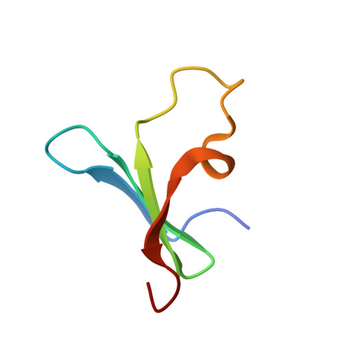
8R3B

Cocrystal form I of the Pent - sulfonato-calix[8]arene complex


Experimental Data Snapshot

  • Method: X-RAY DIFFRACTION
  • Resolution: 1.66 Å
  • R-Value Free: 0.238 
  • R-Value Work: 0.192 
  • R-Value Observed: 0.194 

Starting Model: experimental
View more details

wwPDB Validation   3D Report Full Report


Ligand Structure Quality Assessment 


This is version 1.0 of the entry. See complete history


Literature

Multivalent Calixarene Complexation of a Designed Pentameric Lectin.

Flood, R.J.Cerofolini, L.Fragai, M.Crowley, P.B.

(2024) Biomacromolecules 25: 1303-1309

  • DOI: https://doi.org/10.1021/acs.biomac.3c01280
  • Primary Citation of Related Structures:  
    8R3B, 8R3C, 8R3D

  • PubMed Abstract: 

    We describe complex formation between a designed pentameric β-propeller and the anionic macrocycle sulfonato-calix[8]arene ( sclx 8 ), as characterized by X-ray crystallography and NMR spectroscopy. Two crystal structures and 15 N HSQC experiments reveal a single calixarene binding site in the concave pocket of the β-propeller toroid. Despite the symmetry mismatch between the pentameric protein and the octameric macrocycle, they form a high affinity multivalent complex, with the largest protein-calixarene interface observed to date. This system provides a platform for investigating multivalency.


  • Organizational Affiliation

    SSPC, Science Foundation Ireland Research Centre for Pharmaceuticals, School of Biological and Chemical Sciences, University of Galway, University Road, Galway H91 TK33, Ireland.


Macromolecules
Find similar proteins by:  (by identity cutoff)  |  3D Structure
Entity ID: 1
MoleculeChains Sequence LengthOrganismDetailsImage
Beta propellerA,
B,
C [auth E],
D,
E [auth C]
48Enterobacteria phage L1Mutation(s): 1 
UniProt
Find proteins for A0A140UHM9 (Enterobacteria phage L1)
Explore A0A140UHM9 
Go to UniProtKB:  A0A140UHM9
Entity Groups  
Sequence Clusters30% Identity50% Identity70% Identity90% Identity95% Identity100% Identity
UniProt GroupA0A140UHM9
Sequence Annotations
Expand
  • Reference Sequence
Experimental Data & Validation

Experimental Data

  • Method: X-RAY DIFFRACTION
  • Resolution: 1.66 Å
  • R-Value Free: 0.238 
  • R-Value Work: 0.192 
  • R-Value Observed: 0.194 
  • Space Group: P 43 21 2
Unit Cell:
Length ( Å )Angle ( ˚ )
a = 52.084α = 90
b = 52.084β = 90
c = 177.488γ = 90
Software Package:
Software NamePurpose
PHASERphasing
PHENIXrefinement
autoPROCdata processing
Aimlessdata scaling
XDSdata reduction

Structure Validation

View Full Validation Report


Ligand Structure Quality Assessment 


Entry History & Funding Information

Deposition Data


Funding OrganizationLocationGrant Number
Science Foundation IrelandIreland12/RC/2275_P2

Revision History  (Full details and data files)

  • Version 1.0: 2024-03-06
    Type: Initial release