8R4A

Cryo-EM structure of the D87G lysozyme amyloid fibril


Experimental Data Snapshot

  • Method: ELECTRON MICROSCOPY
  • Resolution: 2.80 Å
  • Aggregation State: HELICAL ARRAY 
  • Reconstruction Method: HELICAL 

wwPDB Validation   3D Report Full Report


This is version 1.1 of the entry. See complete history


Literature

Cryo-EM structure of a lysozyme-derived amyloid fibril from hereditary amyloidosis.

Karimi-Farsijani, S.Sharma, K.Ugrina, M.Kuhn, L.Pfeiffer, P.B.Haupt, C.Wiese, S.Hegenbart, U.Schonland, S.O.Schwierz, N.Schmidt, M.Fandrich, M.

(2024) Nat Commun 15: 9648-9648

  • DOI: https://doi.org/10.1038/s41467-024-54091-7
  • Primary Citation of Related Structures:  
    8R4A

  • PubMed Abstract: 

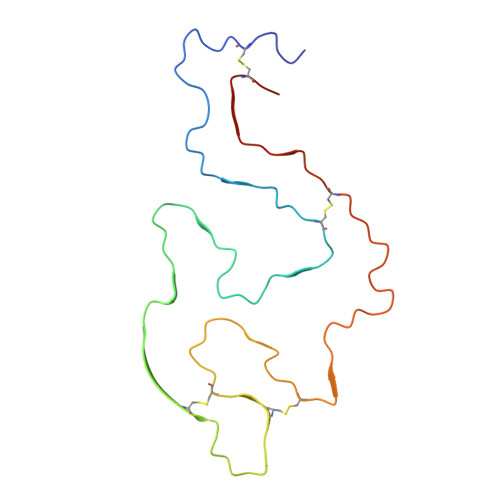
    Systemic ALys amyloidosis is a debilitating protein misfolding disease that arises from the formation of amyloid fibrils from C-type lysozyme. We here present a 2.8 Å cryo-electron microscopy structure of an amyloid fibril, which was isolated from the abdominal fat tissue of a patient who expressed the D87G variant of human lysozyme. We find that the fibril possesses a stable core that is formed by all 130 residues of the fibril precursor protein. There are four disulfide bonds in each fibril protein that connect the same residues as in the globularly folded protein. As the conformation of lysozyme in the fibril is otherwise fundamentally different from native lysozyme, our data provide a structural rationale for the need of protein unfolding in the development of systemic ALys amyloidosis.


  • Organizational Affiliation

    Institute of Protein Biochemistry, Ulm University, Ulm, Germany. sara.karimi-farsijani@uni-ulm.de.


Macromolecules
Find similar proteins by:  (by identity cutoff)  |  3D Structure
Entity ID: 1
MoleculeChains Sequence LengthOrganismDetailsImage
Lysozyme C
A, B, C, D, E
130Homo sapiensMutation(s): 1 
EC: 3.2.1.17
UniProt & NIH Common Fund Data Resources
Find proteins for P61626 (Homo sapiens)
Explore P61626 
Go to UniProtKB:  P61626
PHAROS:  P61626
GTEx:  ENSG00000090382 
Entity Groups  
Sequence Clusters30% Identity50% Identity70% Identity90% Identity95% Identity100% Identity
UniProt GroupP61626
Sequence Annotations
Expand
  • Reference Sequence
Experimental Data & Validation

Experimental Data

  • Method: ELECTRON MICROSCOPY
  • Resolution: 2.80 Å
  • Aggregation State: HELICAL ARRAY 
  • Reconstruction Method: HELICAL 
EM Software:
TaskSoftware PackageVersion
RECONSTRUCTIONRELION
MODEL REFINEMENTCoot

Structure Validation

View Full Validation Report


Entry History & Funding Information

Deposition Data


Funding OrganizationLocationGrant Number
Not funded--

Revision History  (Full details and data files)

  • Version 1.0: 2024-11-20
    Type: Initial release
  • Version 1.1: 2024-11-27
    Changes: Data collection, Database references