8R5V | pdb_00008r5v

Crystal structure of bovine pancreatic ribonuclease A in complex with [Sp-PS]-mU-dT dinucleotide


Experimental Data Snapshot

  • Method: X-RAY DIFFRACTION
  • Resolution: 1.80 Å
  • R-Value Free: 
    0.230 (Depositor), 0.222 (DCC) 
  • R-Value Work: 
    0.164 (Depositor) 

Starting Model: experimental
View more details

wwPDB Validation 3D Report Full Report

Validation slider image for 8R5V

Ligand Structure Quality Assessment 


This is version 1.2 of the entry. See complete history

Literature

P-Stereodefined morpholino dinucleoside 3',5'-phosphorothioates.

Jastrzebska, K.Antonczyk, P.Dolot, R.

(2024) Org Biomol Chem 22: 8737-8742

  • DOI: https://doi.org/10.1039/d4ob01437e
  • Primary Citation Related Structures: 
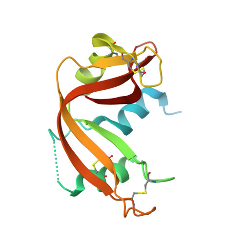
    8R5V

  • PubMed Abstract: 

    Here, we present for the first time the synthesis of P-stereodefined morpholino phosphorothioate analogs by using a modified 1,3,2-oxathiaphospholane method (OTP method) and provide valuable structural insights into their stereochemistry. N -(2-Thio-4,4-pentamethylene-1,3,2-oxathiaphospholane) derivatives of morpholino-type nucleosides ( m U-OTPs) were synthesized, separated into pure P-diastereomers and used to prepare P-stereodefined morpholino dinucleoside 3',5'-phosphorothioates.


  • Organizational Affiliation
    • Centre of Molecular and Macromolecular Studies, Polish Academy of Sciences, Department of Bioorganic Chemistry, Sienkiewicza 112, 90-363 Łódź, Poland. katarzyna.jastrzebska@cbmm.lodz.pl.

Macromolecule Content 

  • Total Structure Weight: 33.77 kDa 
  • Atom Count: 2,413 
  • Modeled Residue Count: 243 
  • Deposited Residue Count: 300 
  • Unique protein chains: 1

Macromolecules

Find similar proteins by:|  3D Structure
Entity ID: 1
MoleculeChains  Sequence LengthOrganismDetailsImage
Ribonuclease pancreatic
A, B
150Bos taurusMutation(s): 0 
EC: 4.6.1.18
UniProt
Find proteins for P61823 (Bos taurus)
Explore P61823 
Go to UniProtKB:  P61823
Entity Groups
Sequence Clusters30% Identity50% Identity70% Identity90% Identity95% Identity100% Identity
UniProt GroupP61823
Sequence Annotations
Expand
Reference Sequence

Small Molecules

Ligands 2 Unique
IDChains Name / Formula / InChI Key2D Diagram3D Interactions
Y4I
(Subject of Investigation/LOI)

Query on Y4I


Download:Ideal Coordinates CCD File
C [auth A]1-[(2~{R},4~{S},5~{R})-5-[[[(2~{R},6~{S})-2-[2,4-bis(oxidanylidene)pyrimidin-1-yl]-6-(hydroxymethyl)morpholin-4-yl]-oxidanyl-phosphinothioyl]oxymethyl]-4-oxidanyl-oxolan-2-yl]-5-methyl-pyrimidine-2,4-dione
C19 H26 N5 O10 P S
JMOYBKKBBMXKAB-OTRGKRLXSA-N
TF4

Query on TF4


Download:Ideal Coordinates CCD File
D [auth A],
E [auth B]
DICHLORO-ACETIC ACID
C2 H2 Cl2 O2
JXTHNDFMNIQAHM-UHFFFAOYSA-N

Experimental Data & Validation

Experimental Data

  • Method: X-RAY DIFFRACTION
  • Resolution: 1.80 Å
  • R-Value Free:  0.230 (Depositor), 0.222 (DCC) 
  • R-Value Work:  0.164 (Depositor) 
Space Group: C 1 2 1
Unit Cell:
Length ( Å )Angle ( ˚ )
a = 100.315α = 90
b = 32.709β = 90.069
c = 72.802γ = 90
Software Package:
Software NamePurpose
REFMACrefinement
CrysalisProdata reduction
Aimlessdata scaling
MOLREPphasing

Structure Validation

View Full Validation Report


Ligand Structure Quality Assessment 


Entry History 

& Funding Information

Deposition Data


Funding OrganizationLocationGrant Number
Polish National Science CentrePoland2021/43/D/ST4/02433

Revision History  (Full details and data files)

  • Version 1.0: 2023-12-06
    Type: Initial release
  • Version 1.1: 2024-10-30
    Changes: Database references, Structure summary
  • Version 1.2: 2024-11-20
    Changes: Database references