An alternative conformation of the N-terminal loop of human dihydroorotate dehydrogenase drives binding to a potent antiproliferative agent.
Alberti, M., Poli, G., Broggini, L., Sainas, S., Rizzi, M., Boschi, D., Ferraris, D.M., Martino, E., Ricagno, S., Tuccinardi, T., Lolli, M.L., Miggiano, R.(2024) Acta Crystallogr D Struct Biol 80: 386-396
- PubMed: 38805244 Search on PubMed
- DOI: https://doi.org/10.1107/S2059798324004066
- Primary Citation Related Structures:
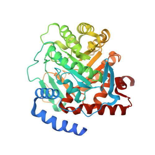
8RAK - PubMed Abstract:
Over the years, human dihydroorotate dehydrogenase (hDHODH), which is a key player in the de novo pyrimidine-biosynthesis pathway, has been targeted in the treatment of several conditions, including autoimmune disorders and acute myelogenous leukaemia, as well as in host-targeted antiviral therapy. A molecular exploration of its inhibitor-binding behaviours yielded promising candidates for innovative drug design. A detailed description of the enzymatic pharmacophore drove the decoration of well-established inhibitory scaffolds, thus gaining further in vitro and in vivo efficacy. In the present work, using X-ray crystallography, an atypical rearrangement was identified in the binding pose of a potent inhibitor characterized by a polar pyridine-based moiety (compound 18). The crystal structure shows that upon binding compound 18 the dynamics of a protein loop involved in a gating mechanism at the cofactor-binding site is modulated by the presence of three water molecules, thus fine-tuning the polarity/hydrophobicity of the binding pocket. These solvent molecules are engaged in the formation of a hydrogen-bond mesh in which one of them establishes a direct contact with the pyridine moiety of compound 18, thus paving the way for a reappraisal of the inhibition of hDHODH. Using an integrated approach, the thermodynamics of such a modulation is described by means of isothermal titration calorimetry coupled with molecular modelling. These structural insights will guide future drug design to obtain a finer K d /logD 7.4 balance and identify membrane-permeable molecules with a drug-like profile in terms of water solubility.
- Department of Pharmaceutical Sciences, University of Piemonte Orientale, Via G. Bovio 6, 28100 Novara, Italy.
Organizational Affiliation: