8RPH

JanthE from Janthinobacterium sp. HH01,ketobutyryl-ThDP


Experimental Data Snapshot

  • Method: X-RAY DIFFRACTION
  • Resolution: 2.96 Å
  • R-Value Free: 0.240 
  • R-Value Work: 0.206 
  • R-Value Observed: 0.208 

Starting Model: in silico
View more details

wwPDB Validation   3D Report Full Report


Ligand Structure Quality Assessment 


This is version 1.3 of the entry. See complete history


Literature

Identification and Characterization of Thiamine Diphosphate-Dependent Lyases with an Unusual CDG Motif.

Lanza, L.Rabe von Pappenheim, F.Bjarnesen, D.Leogrande, C.Paul, A.Krug, L.Tittmann, K.Muller, M.

(2024) Angew Chem Int Ed Engl 63: e202404045-e202404045

  • DOI: https://doi.org/10.1002/anie.202404045
  • Primary Citation of Related Structures:  
    8RPH, 8RPI, 8RPJ

  • PubMed Abstract: 

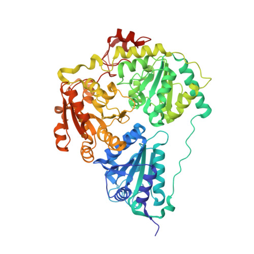
    The thiamine diphosphate (ThDP)-binding motif, characterized by the canonical GDG(X) 24-27 N sequence, is highly conserved among ThDP-dependent enzymes. We investigated a ThDP-dependent lyase (JanthE from Janthinobacterium sp. HH01) with an unusual cysteine (C458) replacing the first glycine of this motif. JanthE exhibits a high substrate promiscuity and accepts long aliphatic α-keto acids as donors. Sterically hindered aromatic aldehydes or non-activated ketones are acceptor substrates, giving access to a variety of secondary and tertiary alcohols as carboligation products. The crystal structure solved at a resolution of 1.9 Å reveals that C458 is not primarily involved in cofactor binding as previously thought for the canonical glycine. Instead, it coordinates methionine 406, thus ensuring the integrity of the active site and the enzyme activity. In addition, we have determined the long-sought genuine tetrahedral intermediates formed with pyruvate and 2-oxobutyrate in the pre-decarboxylation states and deciphered the atomic details for their stabilization in the active site. Collectively, we unravel an unexpected role for the first residue of the ThDP-binding motif and unlock a family of lyases that can perform valuable carboligation reactions.


  • Organizational Affiliation

    Institute of Pharmaceutical Sciences, Albert-Ludwigs-Universität Freiburg, Albertstrasse 25, 79104, Freiburg im Breisgau, Germany.


Macromolecules
Find similar proteins by:  (by identity cutoff)  |  3D Structure
Entity ID: 1
MoleculeChains Sequence LengthOrganismDetailsImage
Thiamine pyrophosphate-binding protein
A, B, C, D, E
A, B, C, D, E, F
619Janthinobacterium sp. HH01Mutation(s): 0 
EC: 2.2.1.6
Entity Groups  
Sequence Clusters30% Identity50% Identity70% Identity90% Identity95% Identity100% Identity
Sequence Annotations
Expand
  • Reference Sequence
Small Molecules
Ligands 4 Unique
IDChains Name / Formula / InChI Key2D Diagram3D Interactions
FAD
Query on FAD

Download Ideal Coordinates CCD File 
G [auth A]
J [auth B]
M [auth C]
P [auth D]
T [auth E]
G [auth A],
J [auth B],
M [auth C],
P [auth D],
T [auth E],
W [auth F]
FLAVIN-ADENINE DINUCLEOTIDE
C27 H33 N9 O15 P2
VWWQXMAJTJZDQX-UYBVJOGSSA-N
A1H2A (Subject of Investigation/LOI)
Query on A1H2A

Download Ideal Coordinates CCD File 
I [auth A]
L [auth B]
O [auth C]
S [auth D]
V [auth E]
I [auth A],
L [auth B],
O [auth C],
S [auth D],
V [auth E],
Y [auth F]
(2~{S})-2-[3-[(4-azanyl-2-methyl-pyrimidin-5-yl)methyl]-4-methyl-5-[2-[oxidanyl(phosphonooxy)phosphoryl]oxyethyl]-1,3-thiazol-2-yl]-2-oxidanyl-butanoic acid
C16 H25 N4 O10 P2 S
FMOBPODOIDIUAY-MRXNPFEDSA-O
PGE
Query on PGE

Download Ideal Coordinates CCD File 
R [auth D]TRIETHYLENE GLYCOL
C6 H14 O4
ZIBGPFATKBEMQZ-UHFFFAOYSA-N
MG
Query on MG

Download Ideal Coordinates CCD File 
H [auth A]
K [auth B]
N [auth C]
Q [auth D]
U [auth E]
H [auth A],
K [auth B],
N [auth C],
Q [auth D],
U [auth E],
X [auth F]
MAGNESIUM ION
Mg
JLVVSXFLKOJNIY-UHFFFAOYSA-N
Experimental Data & Validation

Experimental Data

  • Method: X-RAY DIFFRACTION
  • Resolution: 2.96 Å
  • R-Value Free: 0.240 
  • R-Value Work: 0.206 
  • R-Value Observed: 0.208 
  • Space Group: C 1 2 1
Unit Cell:
Length ( Å )Angle ( ˚ )
a = 173.326α = 90
b = 118.189β = 97.06
c = 201.166γ = 90
Software Package:
Software NamePurpose
PHENIXrefinement
XDSdata reduction
Aimlessdata scaling
PHENIXphasing

Structure Validation

View Full Validation Report


Ligand Structure Quality Assessment 


Entry History & Funding Information

Deposition Data


Funding OrganizationLocationGrant Number
H2020 Marie Curie Actions of the European CommissionEuropean Union956631

Revision History  (Full details and data files)

  • Version 1.0: 2024-06-12
    Type: Initial release
  • Version 1.1: 2024-06-19
    Changes: Database references, Structure summary
  • Version 1.2: 2024-08-07
    Changes: Database references
  • Version 1.3: 2024-08-21
    Changes: Database references