8RTZ

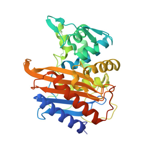
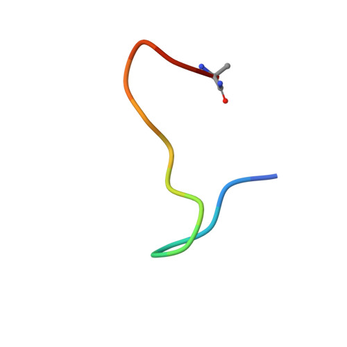
The structure of E. coli penicillin binding protein 3 (PBP3) in complex with a bicyclic peptide inhibitor


Experimental Data Snapshot

  • Method: X-RAY DIFFRACTION
  • Resolution: 1.52 Å
  • R-Value Free: 0.216 
  • R-Value Work: 0.182 

Starting Model: experimental
View more details

wwPDB Validation   3D Report Full Report


Ligand Structure Quality Assessment 


This is version 1.1 of the entry. See complete history


Literature

Discovery and chemical optimisation of a Potent, Bi-cyclic (Bicycle) Antimicrobial Inhibitor of Escherichia coli PBP3

Rowland, C.E.Newman, H.Martin, T.T.Dods, R.Bournakas, N.Wagstaff, J.M.Lewis, N.Stanway, S.J.Blamforth, M.Kessler, C.van Rietschoten, K.Bellini, D.Roper, D.I.Lloyd, A.J.Dowson, C.G.Skynner, M.J.Beswick, P.Dawson, M.

To be published.

Macromolecules
Find similar proteins by:  (by identity cutoff)  |  3D Structure
Entity ID: 1
MoleculeChains Sequence LengthOrganismDetailsImage
Peptidoglycan D,D-transpeptidase FtsIA [auth AAA]348Escherichia coliMutation(s): 0 
Gene Names: ftsIpbpBb0084JW0082
EC: 3.4.16.4
UniProt
Find proteins for P0AD68 (Escherichia coli (strain K12))
Explore P0AD68 
Go to UniProtKB:  P0AD68
Entity Groups  
Sequence Clusters30% Identity50% Identity70% Identity90% Identity95% Identity100% Identity
UniProt GroupP0AD68
Sequence Annotations
Expand
  • Reference Sequence

Find similar proteins by:  Sequence   |   3D Structure  

Entity ID: 2
MoleculeChains Sequence LengthOrganismDetailsImage
Bicyclic peptide inhibitorB [auth A]15synthetic constructMutation(s): 0 
Entity Groups  
Sequence Clusters30% Identity50% Identity70% Identity90% Identity95% Identity100% Identity
Sequence Annotations
Expand
  • Reference Sequence
Small Molecules
Ligands 1 Unique
IDChains Name / Formula / InChI Key2D Diagram3D Interactions
29N (Subject of Investigation/LOI)
Query on 29N

Download Ideal Coordinates CCD File 
C [auth A]1,1',1''-(1,3,5-triazinane-1,3,5-triyl)tripropan-1-one
C12 H21 N3 O3
AEPJNZPJFYDQLM-UHFFFAOYSA-N
Experimental Data & Validation

Experimental Data

  • Method: X-RAY DIFFRACTION
  • Resolution: 1.52 Å
  • R-Value Free: 0.216 
  • R-Value Work: 0.182 
  • Space Group: C 2 2 21
Unit Cell:
Length ( Å )Angle ( ˚ )
a = 97.89α = 90
b = 152.181β = 90
c = 43.675γ = 90
Software Package:
Software NamePurpose
REFMACrefinement
XDSdata reduction
XDSdata scaling
Aimlessdata scaling
PHASERphasing

Structure Validation

View Full Validation Report


Ligand Structure Quality Assessment 


Entry History & Funding Information

Deposition Data


Funding OrganizationLocationGrant Number
Innovate UKUnited Kingdom971626Q1
Innovate UKUnited Kingdom77556
Innovate UKUnited Kingdom82975

Revision History  (Full details and data files)

  • Version 1.0: 2024-04-03
    Type: Initial release
  • Version 1.1: 2024-10-23
    Changes: Structure summary