8SQX | pdb_00008sqx

Solution structure of the basal pilin SpaB from Corynebacterium diphtheriae


Experimental Data Snapshot

  • Method: SOLUTION NMR
  • Conformers Calculated: 200 
  • Conformers Submitted: 20 
  • Selection Criteria: structures with the lowest energy 

wwPDB Validation 3D Report Full Report

Validation slider image for 8SQX

This is version 1.3 of the entry. See complete history

Literature

The basal and major pilins in the Corynebacterium diphtheriae SpaA pilus adopt similar structures that competitively react with the pilin polymerase.

Sue, C.K.Cheung, N.A.Mahoney, B.J.McConnell, S.A.Scully, J.M.Fu, J.Y.Chang, C.Ton-That, H.Loo, J.A.Clubb, R.T.

(2024) Biopolymers 115: e23539-e23539

  • DOI: https://doi.org/10.1002/bip.23539
  • Primary Citation Related Structures: 
    8SQX

  • PubMed Abstract: 

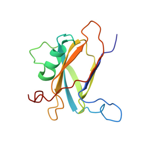
    Many species of pathogenic gram-positive bacteria display covalently crosslinked protein polymers (called pili or fimbriae) that mediate microbial adhesion to host tissues. These structures are assembled by pilus-specific sortase enzymes that join the pilin components together via lysine-isopeptide bonds. The archetypal SpaA pilus from Corynebacterium diphtheriae is built by the Cd SrtA pilus-specific sortase, which crosslinks lysine residues within the SpaA and SpaB pilins to build the shaft and base of the pilus, respectively. Here, we show that Cd SrtA crosslinks SpaB to SpaA via a K139(SpaB)-T494(SpaA) lysine-isopeptide bond. Despite sharing only limited sequence homology, an NMR structure of SpaB reveals striking similarities with the N-terminal domain of SpaA ( N SpaA) that is also crosslinked by Cd SrtA. In particular, both pilins contain similarly positioned reactive lysine residues and adjacent disordered AB loops that are predicted to be involved in the recently proposed "latch" mechanism of isopeptide bond formation. Competition experiments using an inactive SpaB variant and additional NMR studies suggest that SpaB terminates SpaA polymerization by outcompeting N SpaA for access to a shared thioester enzyme-substrate reaction intermediate.


  • Organizational Affiliation
    • Department of Chemistry and Biochemistry, University of California, Los Angeles, California, USA.

Macromolecule Content 

  • Total Structure Weight: 13.61 kDa 
  • Atom Count: 957 
  • Modeled Residue Count: 126 
  • Deposited Residue Count: 126 
  • Unique protein chains: 1

Macromolecules

Find similar proteins by:|  3D Structure
Entity ID: 1
MoleculeChains  Sequence LengthOrganismDetailsImage
Putative surface anchored protein126Corynebacterium diphtheriae NCTC 13129Mutation(s): 0 
Gene Names: DIP2011
UniProt
Find proteins for Q6NF83 (Corynebacterium diphtheriae (strain ATCC 700971 / NCTC 13129 / Biotype gravis))
Explore Q6NF83 
Go to UniProtKB:  Q6NF83
Entity Groups
Sequence Clusters30% Identity50% Identity70% Identity90% Identity95% Identity100% Identity
UniProt GroupQ6NF83
Sequence Annotations
Expand
Reference Sequence

Experimental Data & Validation

Experimental Data

  • Method: SOLUTION NMR
  • Conformers Calculated: 200 
  • Conformers Submitted: 20 
  • Selection Criteria: structures with the lowest energy 

Structure Validation

View Full Validation Report


Entry History 

& Funding Information

Deposition Data


Funding OrganizationLocationGrant Number
National Institutes of Health/National Institute Of Allergy and Infectious Diseases (NIH/NIAID)United StatesAI52217
National Institutes of Health/National Institute of General Medical Sciences (NIH/NIGMS)United StatesT32GM007185

Revision History  (Full details and data files)

  • Version 1.0: 2023-06-07
    Type: Initial release
  • Version 1.1: 2023-06-14
    Changes: Other
  • Version 1.2: 2024-01-31
    Changes: Data collection, Database references
  • Version 1.3: 2024-05-15
    Changes: Database references