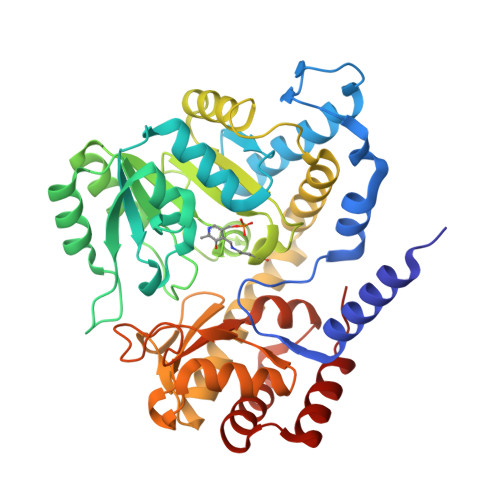
Revealing protonation states and tracking substrate in serine hydroxymethyltransferase with room-temperature X-ray and neutron crystallography.
Drago, V.N., Campos, C., Hooper, M., Collins, A., Gerlits, O., Weiss, K.L., Blakeley, M.P., Phillips, R.S., Kovalevsky, A.(2023) Commun Chem 6: 162-162
- PubMed: 37532884
- DOI: https://doi.org/10.1038/s42004-023-00964-9
- Primary Citation of Related Structures:
8SSJ, 8SSY, 8SUI, 8SUJ - PubMed Abstract:
Pyridoxal 5'-phosphate (PLP)-dependent enzymes utilize a vitamin B 6 -derived cofactor to perform a myriad of chemical transformations on amino acids and other small molecules. Some PLP-dependent enzymes, such as serine hydroxymethyltransferase (SHMT), are promising drug targets for the design of small-molecule antimicrobials and anticancer therapeutics, while others have been used to synthesize pharmaceutical building blocks. Understanding PLP-dependent catalysis and the reaction specificity is crucial to advance structure-assisted drug design and enzyme engineering. Here we report the direct determination of the protonation states in the active site of Thermus thermophilus SHMT (TthSHMT) in the internal aldimine state using room-temperature joint X-ray/neutron crystallography. Conserved active site architecture of the model enzyme TthSHMT and of human mitochondrial SHMT (hSHMT2) were compared by obtaining a room-temperature X-ray structure of hSHMT2, suggesting identical protonation states in the human enzyme. The amino acid substrate serine pathway through the TthSHMT active site cavity was tracked, revealing the peripheral and cationic binding sites that correspond to the pre-Michaelis and pseudo-Michaelis complexes, respectively. At the peripheral binding site, the substrate is bound in the zwitterionic form. By analyzing the observed protonation states, Glu53, but not His residues, is proposed as the general base catalyst, orchestrating the retro-aldol transformation of L-serine into glycine.
- Neutron Scattering Division, Oak Ridge National Laboratory, Oak Ridge, TN, 37831, USA.
Organizational Affiliation:


