8UIQ | pdb_00008uiq

H47Q NicC with 2-mercaptopyridine ligand


Experimental Data Snapshot

  • Method: X-RAY DIFFRACTION
  • Resolution: 2.17 Å
  • R-Value Free: 
    0.283 (Depositor), 0.255 (DCC) 
  • R-Value Work: 
    0.245 (Depositor) 
  • R-Value Observed: 
    0.247 (Depositor) 

Starting Model: experimental
View more details

wwPDB Validation 3D Report Full Report

Validation slider image for 8UIQ

Ligand Structure Quality Assessment 


This is version 1.0 of the entry. See complete history

Literature

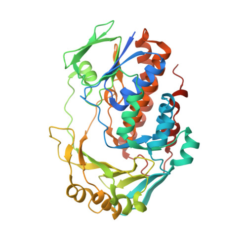
Ligand bound structure of a 6-hydroxynicotinic acid 3-monooxygenase provides mechanistic insights.

Turlington, Z.R.Vaz Ferreira de Macedo, S.Perry, K.Belsky, S.L.Faust, J.A.Snider, M.J.Hicks, K.A.

(2024) Arch Biochem Biophys 752: 109859-109859

  • DOI: https://doi.org/10.1016/j.abb.2023.109859
  • Primary Citation Related Structures: 
    8UIQ, 8UIV

  • PubMed Abstract: 

    6-Hydroxynicotinic acid 3-monooxygenase (NicC) is a bacterial enzyme involved in the degradation of nicotinic acid. This enzyme is a Class A flavin-dependent monooxygenase that catalyzes a unique decarboxylative hydroxylation. The unliganded structure of this enzyme has previously been reported and studied using steady- and transient-state kinetics to support a comprehensive kinetic mechanism. Here we report the crystal structure of the H47Q NicC variant in both a ligand-bound (solved to 2.17 Å resolution) and unliganded (1.51 Å resolution) form. Interestingly, in the liganded form, H47Q NicC is bound to 2-mercaptopyridine (2-MP), a contaminant present in the commercial stock of 6-mercaptopyridine-3-carboxylic acid(6-MNA), a substrate analogue. 2-MP binds weakly to H47Q NicC and is not a substrate for the enzyme. Based on kinetic and thermodynamic characterization, we have fortuitously captured a catalytically inactive H47Q NicC•2-MP complex in our crystal structure. This complex reveals interesting mechanistic details about the reaction catalyzed by 6-hydroxynicotinic acid 3-monooxygenase.


  • Organizational Affiliation
    • Department of Chemistry, State University of New York at Cortland, Cortland, NY, 13045, United States.

Macromolecule Content 

  • Total Structure Weight: 46.32 kDa 
  • Atom Count: 3,012 
  • Modeled Residue Count: 373 
  • Deposited Residue Count: 405 
  • Unique protein chains: 1

Macromolecules

Find similar proteins by:|  3D Structure
Entity ID: 1
MoleculeChains  Sequence LengthOrganismDetailsImage
6-hydroxynicotinate 3-monooxygenase405Pseudomonas putida KT2440Mutation(s): 1 
Gene Names: nicC
EC: 1.14.13.114
UniProt
Find proteins for Q88FY2 (Pseudomonas putida (strain ATCC 47054 / DSM 6125 / CFBP 8728 / NCIMB 11950 / KT2440))
Explore Q88FY2 
Go to UniProtKB:  Q88FY2
Entity Groups
Sequence Clusters30% Identity50% Identity70% Identity90% Identity95% Identity100% Identity
UniProt GroupQ88FY2
Sequence Annotations
Expand
Reference Sequence

Experimental Data & Validation

Experimental Data

  • Method: X-RAY DIFFRACTION
  • Resolution: 2.17 Å
  • R-Value Free:  0.283 (Depositor), 0.255 (DCC) 
  • R-Value Work:  0.245 (Depositor) 
  • R-Value Observed: 0.247 (Depositor) 
Space Group: P 21 21 21
Unit Cell:
Length ( Å )Angle ( ˚ )
a = 47.376α = 90
b = 69.244β = 90
c = 115.27γ = 90
Software Package:
Software NamePurpose
PHENIXrefinement
XDSdata reduction
XDSdata scaling
PHASERphasing

Structure Validation

View Full Validation Report


Ligand Structure Quality Assessment 


Entry History 

& Funding Information

Deposition Data


Funding OrganizationLocationGrant Number
National Science Foundation (NSF, United States)United States1817633

Revision History  (Full details and data files)

  • Version 1.0: 2024-02-21
    Type: Initial release