8UQQ | pdb_00008uqq

Structure of mCLIFY: a circularly permuted yellow fluorescent protein


Experimental Data Snapshot

  • Method: X-RAY DIFFRACTION
  • Resolution: 1.57 Å
  • R-Value Free: 
    0.191 (Depositor), 0.191 (DCC) 
  • R-Value Work: 
    0.179 (Depositor), 0.180 (DCC) 
  • R-Value Observed: 
    0.179 (Depositor) 

Starting Model: experimental
View more details

wwPDB Validation 3D Report Full Report

Validation slider image for 8UQQ

This is version 2.1 of the entry. See complete history

Literature

Characterization and structural basis for the brightness of mCLIFY: A novel monomeric and circularly permuted bright yellow fluorescent protein.

Shweta, H.Gupta, K.Zhou, Y.Cui, X.Li, S.Lu, Z.Goldman, Y.E.Dantzig, J.A.

(2025) Biophys J 124: 2133-2149

  • DOI: https://doi.org/10.1016/j.bpj.2025.05.012
  • Primary Citation Related Structures: 
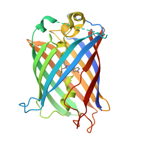
    8UQQ

  • PubMed Abstract: 

    Ongoing improvements of genetically encoded fluorescent proteins have enhanced cellular localization studies and performance of biosensors, such as environmentally or mechanically sensitive fluorescence resonance energy transfer pairs, in cell biological and biophysical research. The brightest yellow fluorescent protein, widely used in these studies is YPet, derived from the jellyfish Aequorea victoria via the GFP derivative Venus. YPet dimerizes at concentrations used in cellular studies (K D 1-2 = 3.4 μM) which impacts quantitative interpretation of emission intensity, rotational freedom, energy transfer, and lifetime. Although YPet is nearly 30% brighter than Venus, no atomic structures of YPet have been reported to ascertain the structural differences leading to the higher brightness, possibly due to the tendency to dimerize or oligomerize. Here, we report properties of a new YPet derivative, mCLIFY, a monomeric, bright, yellow, and long-lived fluorescent protein created by circular permutation of YPet and substitution of the amino acid residues thought to mediate dimerization. mCLIFY retains the advantageous photophysical properties of YPet but does not dimerize at least up to 40 μM concentration. We determined the atomic structure of mCLIFY at 1.57-Å resolution. Extensive characterization of the photophysical and structural properties of YPet and mCLIFY allowed us to elucidate the bases of their long lifetimes, enhanced brightness, and the difference in propensity to dimerize.


  • Organizational Affiliation
    • Pennsylvania Muscle Institute, Perelman School of Medicine, University of Pennsylvania, Philadelphia, Pennsylvania, USA; Center for Engineering Mechanobiology (CEMB), Perelman School of Medicine, University of Pennsylvania, Philadelphia, Pennsylvania, USA; Department of Physiology, Perelman School of Medicine, University of Pennsylvania, Philadelphia, Pennsylvania, USA; Department of Pharmacology and of Cellular and Molecular Biology, University of California at Davis, Davis, California, USA.

Macromolecule Content 

  • Total Structure Weight: 28.17 kDa 
  • Atom Count: 1,942 
  • Modeled Residue Count: 224 
  • Deposited Residue Count: 251 
  • Unique protein chains: 1

Macromolecules

Find similar proteins by:|  3D Structure
Entity ID: 1
MoleculeChains  Sequence LengthOrganismDetailsImage
mCLIFY251Aequorea victoriaMutation(s): 0 
UniProt
Find proteins for P42212 (Aequorea victoria)
Explore P42212 
Go to UniProtKB:  P42212
Entity Groups
Sequence Clusters30% Identity50% Identity70% Identity90% Identity95% Identity100% Identity
UniProt GroupP42212
Sequence Annotations
Expand
Reference Sequence

Small Molecules

Modified Residues  1 Unique
IDChains TypeFormula2D DiagramParent
CR2
Query on CR2
A
L-PEPTIDE LINKINGC13 H13 N3 O4GLY, TYR, GLY

Experimental Data & Validation

Experimental Data

  • Method: X-RAY DIFFRACTION
  • Resolution: 1.57 Å
  • R-Value Free:  0.191 (Depositor), 0.191 (DCC) 
  • R-Value Work:  0.179 (Depositor), 0.180 (DCC) 
  • R-Value Observed: 0.179 (Depositor) 
Space Group: P 2 21 21
Unit Cell:
Length ( Å )Angle ( ˚ )
a = 35.02α = 90
b = 69.956β = 90
c = 84.671γ = 90
Software Package:
Software NamePurpose
PHENIXrefinement
MOSFLMdata reduction
SCALAdata scaling
PHENIXphasing

Structure Validation

View Full Validation Report


Entry History 

& Funding Information

Deposition Data


Funding OrganizationLocationGrant Number
National Institutes of Health/National Institute of General Medical Sciences (NIH/NIGMS)United StatesR35-GM118139
National Institutes of Health/National Institute of Diabetes and Digestive and Kidney Disease (NIH/NIDDK)United StatesDK125521
National Institutes of Health/National Institute of General Medical Sciences (NIH/NIGMS)United StatesGM055560
Other governmentUnited StatesNIH S10 instrumentation grant: S10OD021573
National Science Foundation (NSF, United States)United StatesCMMI: 15-48571
Other privateUnited StatesJohnson Research Foundation
Other governmentUnited StatesNIH S10 instrumentation grant: S10OD030413

Revision History  (Full details and data files)

  • Version 1.0: 2024-10-30
    Type: Initial release
  • Version 2.0: 2026-03-18
    Changes: Polymer sequence
  • Version 2.1: 2026-05-13
    Changes: Database references