


Discovery of amentoflavone as a natural PDE4 inhibitor with anti-fibrotic effects
Chen, Z., Shi, Y., Zhong, F., Zhang, K., Zhang, F., Xie, S., Cheng, Z., Zhou, Q., Huang, Y.Y., Luo, H.B.(2024) Chin Chem Lett : 109956
Experimental Data Snapshot
Starting Model: experimental
View more details
Entity ID: 1 | |||||
|---|---|---|---|---|---|
| Molecule | Chains | Sequence Length | Organism | Details | Image |
| 3',5'-cyclic-AMP phosphodiesterase 4D | 506 | Homo sapiens | Mutation(s): 0 Gene Names: PDE4D, DPDE3 EC: 3.1.4.53 | 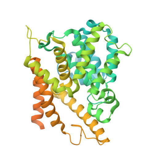 | |
UniProt & NIH Common Fund Data Resources | |||||
PHAROS: Q08499 GTEx: ENSG00000113448 | |||||
Entity Groups | |||||
| Sequence Clusters | 30% Identity50% Identity70% Identity90% Identity95% Identity100% Identity | ||||
| UniProt Group | Q08499 | ||||
Sequence AnnotationsExpand | |||||
Reference Sequence | |||||
| Ligands 3 Unique | |||||
|---|---|---|---|---|---|
| ID | Chains | Name / Formula / InChI Key | 2D Diagram | 3D Interactions | |
| A1D6Q (Subject of Investigation/LOI) Download:Ideal Coordinates CCD File | E [auth A], H [auth B] | 8-[5-[5,7-bis(oxidanyl)-4-oxidanylidene-chromen-2-yl]-2-oxidanyl-phenyl]-2-(4-hydroxyphenyl)-5,7-bis(oxidanyl)chromen-4-one C30 H18 O10 YUSWMAULDXZHPY-UHFFFAOYSA-N |  | ||
| ZN Download:Ideal Coordinates CCD File | C [auth A], F [auth B] | ZINC ION Zn PTFCDOFLOPIGGS-UHFFFAOYSA-N |  | ||
| MG Download:Ideal Coordinates CCD File | D [auth A], G [auth B] | MAGNESIUM ION Mg JLVVSXFLKOJNIY-UHFFFAOYSA-N |  | ||
| Length ( Å ) | Angle ( ˚ ) |
|---|---|
| a = 57.901 | α = 90 |
| b = 78.791 | β = 90 |
| c = 162.394 | γ = 90 |
| Software Name | Purpose |
|---|---|
| REFMAC | refinement |
| PHENIX | refinement |
| Funding Organization | Location | Grant Number |
|---|---|---|
| National Natural Science Foundation of China (NSFC) | China | 22277019 |


