Structure of a fungal LPMO bound to ligands
Banerjee, S., Huang, Z., Brander, S., Johansen, K.S., Lo Leggio, L.To be published.
Experimental Data Snapshot
Entity ID: 1 | |||||
|---|---|---|---|---|---|
| Molecule | Chains | Sequence Length | Organism | Details | Image |
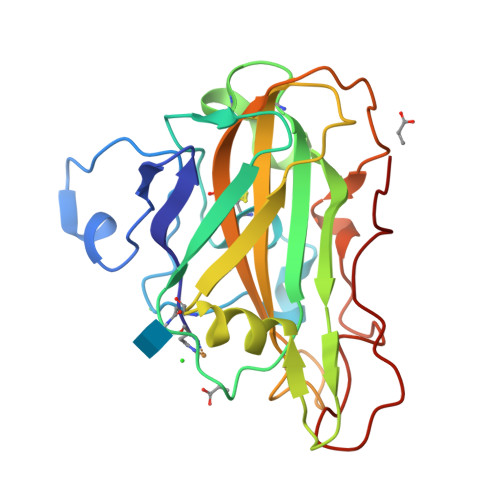
| Gh61 isozyme a | A [auth AAA] | 228 | Thermoascus aurantiacus | Mutation(s): 0 EC: 3.2.1.4 (PDB Primary Data), 1.14.99.56 (UniProt) |  |
UniProt | |||||
Find proteins for G3XAP7 (Thermoascus aurantiacus) Explore G3XAP7 Go to UniProtKB: G3XAP7 | |||||
Entity Groups | |||||
| Sequence Clusters | 30% Identity50% Identity70% Identity90% Identity95% Identity100% Identity | ||||
| UniProt Group | G3XAP7 | ||||
Glycosylation | |||||
| Glycosylation Sites: 1 | |||||
Sequence AnnotationsExpand | |||||
| |||||
| Ligands 5 Unique | |||||
|---|---|---|---|---|---|
| ID | Chains | Name / Formula / InChI Key | 2D Diagram | 3D Interactions | |
| NAG Query on NAG | C [auth AAA] | 2-acetamido-2-deoxy-beta-D-glucopyranose C8 H15 N O6 OVRNDRQMDRJTHS-FMDGEEDCSA-N |  | ||
| 2HA (Subject of Investigation/LOI) Query on 2HA | E [auth AAA] | Dihydroxyacetone C3 H6 O3 RXKJFZQQPQGTFL-UHFFFAOYSA-N |  | ||
| AKR Query on AKR | D [auth AAA], F [auth AAA] | ACRYLIC ACID C3 H4 O2 NIXOWILDQLNWCW-UHFFFAOYSA-N |  | ||
| CU Query on CU | B [auth AAA] | COPPER (II) ION Cu JPVYNHNXODAKFH-UHFFFAOYSA-N |  | ||
| CL Query on CL | G [auth AAA], H [auth AAA] | CHLORIDE ION Cl VEXZGXHMUGYJMC-UHFFFAOYSA-M |  | ||
| Modified Residues 1 Unique | |||||
|---|---|---|---|---|---|
| ID | Chains | Type | Formula | 2D Diagram | Parent |
| HIC Query on HIC | A [auth AAA] | L-PEPTIDE LINKING | C7 H11 N3 O2 |  | HIS |
| Length ( Å ) | Angle ( ˚ ) |
|---|---|
| a = 34.402 | α = 90 |
| b = 87.312 | β = 104.812 |
| c = 37.482 | γ = 90 |
| Software Name | Purpose |
|---|---|
| REFMAC | refinement |
| XDS | data reduction |
| Aimless | data scaling |
| REFMAC | phasing |
| Funding Organization | Location | Grant Number |
|---|---|---|
| Novo Nordisk Foundation | Denmark | -- |
| Danish Council for Independent Research | Denmark | -- |
| Swedish Research Council | Sweden | -- |
RCSB PDB is hosted by

RCSB PDB is a member of the

All questions and comments will receive a response in a timely manner.