8GBE

Structure of a viral gasdermin protein A47 from Eptesipox virus


Experimental Data Snapshot

  • Method: X-RAY DIFFRACTION
  • Resolution: 1.46 Å
  • R-Value Free: 0.212 
  • R-Value Work: 0.191 
  • R-Value Observed: 0.192 

wwPDB Validation   3D Report Full Report


This is version 1.1 of the entry. See complete history


Literature

Structural homology screens reveal poxvirus-encoded proteins impacting inflammasome-mediated defenses.

Boys, I.N.Johnson, A.G.Quinlan, M.Kranzusch, P.J.Elde, N.C.

(2023) Biorxiv 

  • DOI: https://doi.org/10.1101/2023.02.26.529821
  • Primary Citation of Related Structures:  
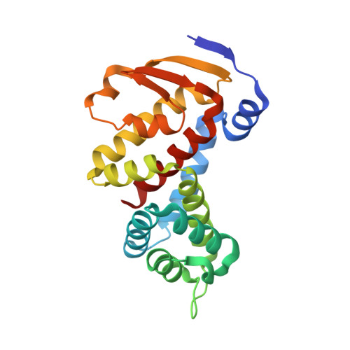
    8GBE

  • PubMed Abstract: 

    Viruses acquire host genes via horizontal gene transfer and can express them to manipulate host biology during infections. Some viral and host homologs retain sequence identity, but evolutionary divergence can obscure host origins. We used structural modeling to compare vaccinia virus proteins with metazoan proteomes. We identified vaccinia A47L as a homolog of gasdermins, the executioners of pyroptosis. An X-ray crystal structure of A47 confirmed this homology and cell-based assays revealed that A47 inhibits pyroptosis. We also identified vaccinia C1L as the product of a cryptic gene fusion event coupling a Bcl-2 related fold with a pyrin domain. C1 associates with components of the inflammasome, a cytosolic innate immune sensor involved in pyroptosis, yet paradoxically enhances inflammasome activity, suggesting a benefit to poxvirus replication in some circumstances. Our findings demonstrate the potential of structural homology screens to reveal genes that viruses capture from hosts and repurpose to benefit viral fitness.


  • Organizational Affiliation

    Department of Human Genetics, University of Utah, Salt Lake City, Utah, 84112 USA.


Macromolecules
Find similar proteins by:  (by identity cutoff)  |  3D Structure
Entity ID: 1
MoleculeChains Sequence LengthOrganismDetailsImage
Protein A47207Eptesipox virusMutation(s): 0 
Gene Names: EPTV-WA-146
UniProt
Find proteins for A0A220T6L2 (Eptesipox virus)
Explore A0A220T6L2 
Go to UniProtKB:  A0A220T6L2
Entity Groups  
Sequence Clusters30% Identity50% Identity70% Identity90% Identity95% Identity100% Identity
UniProt GroupA0A220T6L2
Sequence Annotations
Expand
  • Reference Sequence
Experimental Data & Validation

Experimental Data

  • Method: X-RAY DIFFRACTION
  • Resolution: 1.46 Å
  • R-Value Free: 0.212 
  • R-Value Work: 0.191 
  • R-Value Observed: 0.192 
  • Space Group: C 2 2 21
Unit Cell:
Length ( Å )Angle ( ˚ )
a = 64.465α = 90
b = 74.14β = 90
c = 95.599γ = 90
Software Package:
Software NamePurpose
PHENIXrefinement
XDSdata reduction
Aimlessdata scaling
PHENIXphasing

Structure Validation

View Full Validation Report


Entry History & Funding Information

Deposition Data


Funding OrganizationLocationGrant Number
National Institutes of Health/National Institute of General Medical Sciences (NIH/NIGMS)United States--

Revision History  (Full details and data files)

  • Version 1.0: 2023-03-15
    Type: Initial release
  • Version 1.1: 2023-03-22
    Changes: Database references