8R3B | pdb_00008r3b

Cocrystal form I of the Pent - sulfonato-calix[8]arene complex


Experimental Data Snapshot

  • Method: X-RAY DIFFRACTION
  • Resolution: 1.66 Å
  • R-Value Free: 
    0.238 (Depositor), 0.230 (DCC) 
  • R-Value Work: 
    0.192 (Depositor), 0.190 (DCC) 
  • R-Value Observed: 
    0.194 (Depositor) 

Starting Model: experimental
View more details

wwPDB Validation   3D Report Full Report


Ligand Structure Quality Assessment 

Created with Raphaël 2.3.0Worse 01 BetterLigand structure goodness of fit to experimental dataBest fitted EVBClick on this verticalbar to view details

This is version 1.0 of the entry. See complete history


Literature

Multivalent Calixarene Complexation of a Designed Pentameric Lectin.

Flood, R.J.Cerofolini, L.Fragai, M.Crowley, P.B.

(2024) Biomacromolecules 25: 1303-1309

  • DOI: https://doi.org/10.1021/acs.biomac.3c01280
  • Primary Citation of Related Structures:  
    8R3B, 8R3C, 8R3D

  • PubMed Abstract: 

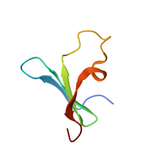
    We describe complex formation between a designed pentameric β-propeller and the anionic macrocycle sulfonato-calix[8]arene ( sclx 8 ), as characterized by X-ray crystallography and NMR spectroscopy. Two crystal structures and 15 N HSQC experiments reveal a single calixarene binding site in the concave pocket of the β-propeller toroid. Despite the symmetry mismatch between the pentameric protein and the octameric macrocycle, they form a high affinity multivalent complex, with the largest protein-calixarene interface observed to date. This system provides a platform for investigating multivalency.


  • Organizational Affiliation

    SSPC, Science Foundation Ireland Research Centre for Pharmaceuticals, School of Biological and Chemical Sciences, University of Galway, University Road, Galway H91 TK33, Ireland.


Macromolecules
Find similar proteins by:  (by identity cutoff)  |  3D Structure
Entity ID: 1
MoleculeChains Sequence LengthOrganismDetailsImage
Beta propellerA,
B,
C [auth E],
D,
E [auth C]
48Enterobacteria phage L1Mutation(s): 1 
UniProt
Find proteins for A0A140UHM9 (Enterobacteria phage L1)
Explore A0A140UHM9 
Go to UniProtKB:  A0A140UHM9
Entity Groups  
Sequence Clusters30% Identity50% Identity70% Identity90% Identity95% Identity100% Identity
UniProt GroupA0A140UHM9
Sequence Annotations
Expand
  • Reference Sequence
Experimental Data & Validation

Experimental Data

  • Method: X-RAY DIFFRACTION
  • Resolution: 1.66 Å
  • R-Value Free:  0.238 (Depositor), 0.230 (DCC) 
  • R-Value Work:  0.192 (Depositor), 0.190 (DCC) 
  • R-Value Observed: 0.194 (Depositor) 
Space Group: P 43 21 2
Unit Cell:
Length ( Å )Angle ( ˚ )
a = 52.084α = 90
b = 52.084β = 90
c = 177.488γ = 90
Software Package:
Software NamePurpose
PHASERphasing
PHENIXrefinement
autoPROCdata processing
Aimlessdata scaling
XDSdata reduction

Structure Validation

View Full Validation Report


Ligand Structure Quality Assessment 

Created with Raphaël 2.3.0Worse 01 BetterLigand structure goodness of fit to experimental dataBest fitted EVBClick on this verticalbar to view details

Entry History & Funding Information

Deposition Data


Funding OrganizationLocationGrant Number
Science Foundation IrelandIreland12/RC/2275_P2

Revision History  (Full details and data files)

  • Version 1.0: 2024-03-06
    Type: Initial release