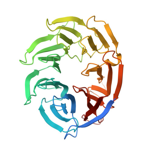
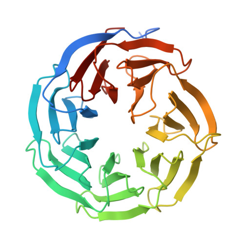
9B9H

Crystal structure of the ternary complex of DCAF1 and WDR5 with PROTAC, OICR-40333


Experimental Data Snapshot

  • Method: X-RAY DIFFRACTION
  • Resolution: 2.06 Å
  • R-Value Free: 0.229 
  • R-Value Work: 0.211 
  • R-Value Observed: 0.212 

Starting Models: experimental
View more details

wwPDB Validation   3D Report Full Report


Ligand Structure Quality Assessment 


This is version 1.1 of the entry. See complete history


Literature

Crystal structures of DCAF1-PROTAC-WDR5 ternary complexes provide insight into DCAF1 substrate specificity.

Mabanglo, M.F.Wilson, B.Noureldin, M.Kimani, S.W.Mamai, A.Krausser, C.Gonzalez-Alvarez, H.Srivastava, S.Mohammed, M.Hoffer, L.Chan, M.Avrumutsoae, J.Li, A.S.M.Hajian, T.Tucker, S.Green, S.Szewczyk, M.Barsyte-Lovejoy, D.Santhakumar, V.Ackloo, S.Loppnau, P.Li, Y.Seitova, A.Kiyota, T.Wang, J.G.Prive, G.G.Kuntz, D.A.Patel, B.Rathod, V.Vala, A.Rout, B.Aman, A.Poda, G.Uehling, D.Ramnauth, J.Halabelian, L.Marcellus, R.Al-Awar, R.Vedadi, M.

(2024) Nat Commun 15: 10165-10165

  • DOI: https://doi.org/10.1038/s41467-024-54500-x
  • Primary Citation of Related Structures:  
    9B9H, 9B9T, 9B9W, 9BA2, 9DLW

  • PubMed Abstract: 

    Proteolysis-targeting chimeras (PROTACs) have been explored for the degradation of drug targets for more than two decades. However, only a handful of E3 ligase substrate receptors have been efficiently used. Downregulation and mutation of these receptors would reduce the effectiveness of such PROTACs. We recently developed potent ligands for DCAF1, a substrate receptor of EDVP and CUL4 E3 ligases. Here, we focus on DCAF1 toward the development of PROTACs for WDR5, a drug target in various cancers. We report four DCAF1-based PROTACs with endogenous and exogenous WDR5 degradation effects and high-resolution crystal structures of the ternary complexes of DCAF1-PROTAC-WDR5. The structures reveal detailed insights into the interaction of DCAF1 with various WDR5-PROTACs, indicating a significant role of DCAF1 loops in providing needed surface plasticity, and reflecting the mechanism by which DCAF1 functions as a substrate receptor for E3 ligases with diverse sets of substrates.


  • Organizational Affiliation

    Drug Discovery Program, Ontario Institute for Cancer Research, Toronto, ON, Canada.


Macromolecules
Find similar proteins by:  (by identity cutoff)  |  3D Structure
Entity ID: 1
MoleculeChains Sequence LengthOrganismDetailsImage
DDB1- and CUL4-associated factor 1A [auth B]338Homo sapiensMutation(s): 0 
Gene Names: DCAF1KIAA0800RIPVPRBP
EC: 2.7.11.1
UniProt & NIH Common Fund Data Resources
Find proteins for Q9Y4B6 (Homo sapiens)
Explore Q9Y4B6 
Go to UniProtKB:  Q9Y4B6
PHAROS:  Q9Y4B6
GTEx:  ENSG00000145041 
Entity Groups  
Sequence Clusters30% Identity50% Identity70% Identity90% Identity95% Identity100% Identity
UniProt GroupQ9Y4B6
Sequence Annotations
Expand
  • Reference Sequence
Find similar proteins by:  (by identity cutoff)  |  3D Structure
Entity ID: 2
MoleculeChains Sequence LengthOrganismDetailsImage
WD repeat-containing protein 5B [auth A]329Homo sapiensMutation(s): 0 
Gene Names: WDR5BIG3
UniProt & NIH Common Fund Data Resources
Find proteins for P61964 (Homo sapiens)
Explore P61964 
Go to UniProtKB:  P61964
PHAROS:  P61964
GTEx:  ENSG00000196363 
Entity Groups  
Sequence Clusters30% Identity50% Identity70% Identity90% Identity95% Identity100% Identity
UniProt GroupP61964
Sequence Annotations
Expand
  • Reference Sequence
Small Molecules
Ligands 1 Unique
IDChains Name / Formula / InChI Key2D Diagram3D Interactions
A1AM2 (Subject of Investigation/LOI)
Query on A1AM2

Download Ideal Coordinates CCD File 
C [auth A]N-{(1P)-5'-({(17E)-18-[(3P)-4-{[(1S)-3-amino-1-(3-chloro-4-fluorophenyl)-3-oxopropyl]carbamoyl}-3-(4-chloro-2-fluorophenyl)-1H-pyrrol-2-yl]-16-oxo-3,6,9,12-tetraoxa-15-azaoctadec-17-en-1-yl}carbamoyl)-2'-fluoro-4-[(3R,5S)-3,4,5-trimethylpiperazin-1-yl][1,1'-biphenyl]-3-yl}-6-oxo-4-(trifluoromethyl)-1,6-dihydropyridine-3-carboxamide
C60 H63 Cl2 F6 N9 O10
GSAYJJSBWPSRLK-KXTAGSPYSA-N
Experimental Data & Validation

Experimental Data

  • Method: X-RAY DIFFRACTION
  • Resolution: 2.06 Å
  • R-Value Free: 0.229 
  • R-Value Work: 0.211 
  • R-Value Observed: 0.212 
  • Space Group: P 21 21 21
Unit Cell:
Length ( Å )Angle ( ˚ )
a = 84.213α = 90
b = 85.143β = 90
c = 111.644γ = 90
Software Package:
Software NamePurpose
PHENIXrefinement
XDSdata reduction
pointlessdata scaling
PHASERphasing

Structure Validation

View Full Validation Report


Ligand Structure Quality Assessment 


Entry History & Funding Information

Deposition Data


Funding OrganizationLocationGrant Number
Ontario Institute for Cancer ResearchCanada--

Revision History  (Full details and data files)

  • Version 1.0: 2024-11-06
    Type: Initial release
  • Version 1.1: 2024-12-04
    Changes: Database references