9ENI | pdb_00009eni

L-amino acid oxidase 4 (HcLAAO4) from the fungus Hebeloma cylindrosporum in complex with L-glutamine


Experimental Data Snapshot

  • Method: X-RAY DIFFRACTION
  • Resolution: 2.10 Å
  • R-Value Free: 
    0.203 (Depositor), 0.203 (DCC) 
  • R-Value Work: 
    0.172 (Depositor), 0.171 (DCC) 
  • R-Value Observed: 
    0.173 (Depositor) 

Starting Model: experimental
View more details

wwPDB Validation 3D Report Full Report

Validation slider image for 9ENI

Ligand Structure Quality Assessment 


This is version 1.2 of the entry. See complete history

Literature

Crystal structure and enzyme engineering of the broad substrate spectrum l-amino acid oxidase 4 from the fungus Hebeloma cylindrosporum.

Koopmeiners, S.Gilzer, D.Widmann, C.Berelsmann, N.Spross, J.Niemann, H.H.Fischer von Mollard, G.

(2024) FEBS Lett 598: 2306-2320

  • DOI: https://doi.org/10.1002/1873-3468.15002
  • Primary Citation Related Structures: 
    9ENH, 9ENI, 9ENJ, 9ENK, 9ENN

  • PubMed Abstract: 

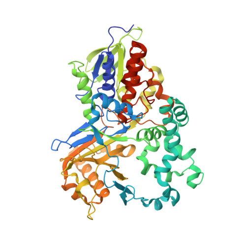
    l-Amino acid oxidases (LAAOs) catalyze the oxidative deamination of l-amino acids to α-keto acids. Recombinant production of LAAOs with broad substrate spectrum remains a formidable challenge. We previously achieved this for the highly active and thermostable LAAO4 of Hebeloma cylindrosporum (HcLAAO4). Here, we crystallized a proteolytically truncated surface entropy reduction variant of HcLAAO4 and solved its structure in substrate-free form and in complex with diverse substrates. The ability to support the aliphatic portion of a substrate's side chain by an overall hydrophobic active site is responsible for the broad substrate spectrum of HcLAAO4, including l-amino acids with big aromatic, acidic and basic side chains. Based on the structural findings, we generated an E288H variant with increased activity toward pharmaceutical building blocks of high interest.


  • Organizational Affiliation
    • Biochemistry III, Department of Chemistry, Bielefeld University, Bielefeld, Germany.

Macromolecule Content 

  • Total Structure Weight: 257.76 kDa 
  • Atom Count: 18,620 
  • Modeled Residue Count: 2,135 
  • Deposited Residue Count: 2,248 
  • Unique protein chains: 1

Macromolecules

Find similar proteins by:|  3D Structure
Entity ID: 1
MoleculeChains  Sequence LengthOrganismDetailsImage
L-amino acid oxidase 4
A, B, C, D
562Hebeloma cylindrosporumMutation(s): 2 
EC: 1.4.3.2 (PDB Primary Data), 1.4.3.25 (UniProt), 1.4.3.11 (UniProt), 1.4.3.14 (UniProt), 1.4.3 (UniProt)
UniProt
Find proteins for S4S6Z0 (Hebeloma cylindrosporum)
Explore S4S6Z0 
Go to UniProtKB:  S4S6Z0
Entity Groups
Sequence Clusters30% Identity50% Identity70% Identity90% Identity95% Identity100% Identity
UniProt GroupS4S6Z0
Sequence Annotations
Expand
Reference Sequence

Small Molecules

Ligands 5 Unique
IDChains Name / Formula / InChI Key2D Diagram3D Interactions
FDA
(Subject of Investigation/LOI)

Query on FDA


Download:Ideal Coordinates CCD File
DB [auth D],
E [auth A],
OA [auth C],
Y [auth B]
DIHYDROFLAVINE-ADENINE DINUCLEOTIDE
C27 H35 N9 O15 P2
YPZRHBJKEMOYQH-UYBVJOGSSA-N
GLN
(Subject of Investigation/LOI)

Query on GLN


Download:Ideal Coordinates CCD File
EB [auth D],
F [auth A],
PA [auth C],
Z [auth B]
GLUTAMINE
C5 H10 N2 O3
ZDXPYRJPNDTMRX-VKHMYHEASA-N
SO4

Query on SO4


Download:Ideal Coordinates CCD File
AA [auth B]
BA [auth B]
CA [auth B]
DA [auth B]
EA [auth B]
AA [auth B],
BA [auth B],
CA [auth B],
DA [auth B],
EA [auth B],
FA [auth B],
FB [auth D],
G [auth A],
GA [auth B],
GB [auth D],
H [auth A],
HA [auth B],
HB [auth D],
I [auth A],
IA [auth B],
IB [auth D],
J [auth A],
JA [auth B],
JB [auth D],
K [auth A],
KA [auth B],
KB [auth D],
L [auth A],
LB [auth D],
M [auth A],
MB [auth D],
N [auth A],
NB [auth D],
O [auth A],
OB [auth D],
P [auth A],
PB [auth D],
Q [auth A],
QA [auth C],
R [auth A],
RA [auth C],
S [auth A],
SA [auth C],
TA [auth C],
UA [auth C],
VA [auth C],
WA [auth C],
XA [auth C],
YA [auth C],
ZA [auth C]
SULFATE ION
O4 S
QAOWNCQODCNURD-UHFFFAOYSA-L
PGO

Query on PGO


Download:Ideal Coordinates CCD File
LA [auth B],
V [auth A]
S-1,2-PROPANEDIOL
C3 H8 O2
DNIAPMSPPWPWGF-VKHMYHEASA-N
PGR

Query on PGR


Download:Ideal Coordinates CCD File
AB [auth C]
BB [auth C]
CB [auth C]
MA [auth B]
NA [auth B]
AB [auth C],
BB [auth C],
CB [auth C],
MA [auth B],
NA [auth B],
QB [auth D],
T [auth A],
U [auth A],
W [auth A],
X [auth A]
R-1,2-PROPANEDIOL
C3 H8 O2
DNIAPMSPPWPWGF-GSVOUGTGSA-N

Experimental Data & Validation

Experimental Data

  • Method: X-RAY DIFFRACTION
  • Resolution: 2.10 Å
  • R-Value Free:  0.203 (Depositor), 0.203 (DCC) 
  • R-Value Work:  0.172 (Depositor), 0.171 (DCC) 
  • R-Value Observed: 0.173 (Depositor) 
Space Group: P 1 21 1
Unit Cell:
Length ( Å )Angle ( ˚ )
a = 84.71α = 90
b = 131.74β = 95.949
c = 107.25γ = 90
Software Package:
Software NamePurpose
PHENIXrefinement
XDSdata reduction
XSCALEdata scaling
PHASERphasing

Structure Validation

View Full Validation Report


Ligand Structure Quality Assessment 


Entry History 

& Funding Information

Deposition Data


Funding OrganizationLocationGrant Number
Not funded--

Revision History  (Full details and data files)

  • Version 1.0: 2024-08-14
    Type: Initial release
  • Version 1.1: 2024-08-28
    Changes: Database references
  • Version 1.2: 2024-10-09
    Changes: Database references, Structure summary