9FWW

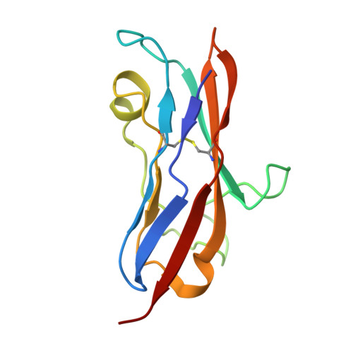
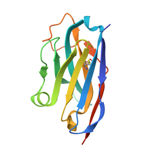
Human NKp30 in complex with a VHH variant


Experimental Data Snapshot

  • Method: X-RAY DIFFRACTION
  • Resolution: 1.84 Å
  • R-Value Free: 0.208 
  • R-Value Work: 0.199 
  • R-Value Observed: 0.200 

Starting Model: experimental
View more details

wwPDB Validation   3D Report Full Report


Ligand Structure Quality Assessment 


This is version 1.0 of the entry. See complete history


Literature

On the humanization of VHHs: Prospective case studies, experimental and computational characterization of structural determinants for functionality.

Fernandez-Quintero, M.L.Guarnera, E.Musil, D.Pekar, L.Sellmann, C.Freire, F.Sousa, R.L.Santos, S.P.Freitas, M.C.Bandeiras, T.M.Silva, M.M.S.Loeffler, J.R.Ward, A.B.Harwardt, J.Zielonka, S.Evers, A.

(2024) Protein Sci 33: e5176-e5176

  • DOI: https://doi.org/10.1002/pro.5176
  • Primary Citation of Related Structures:  
    9FWW, 9FXF

  • PubMed Abstract: 

    The humanization of camelid-derived variable domain heavy chain antibodies (VHHs) poses challenges including immunogenicity, stability, and potential reduction of affinity. Critical to this process are complementarity-determining regions (CDRs), Vernier and Hallmark residues, shaping the three-dimensional fold and influencing VHH structure and function. Additionally, the presence of non-canonical disulfide bonds further contributes to conformational stability and antigen binding. In this study, we systematically humanized two camelid-derived VHHs targeting the natural cytotoxicity receptor NKp30. Key structural positions in Vernier and Hallmark regions were exchanged with residues from the most similar human germline sequences. The resulting variants were characterized for binding affinities, yield, and purity. Structural binding modes were elucidated through crystal structure determination and AlphaFold2 predictions, providing insights into differences in binding affinity. Comparative structural and molecular dynamics characterizations of selected variants were performed to rationalize their functional properties and elucidate the role of specific sequence motifs in antigen binding. Furthermore, systematic analyses of next-generation sequencing (NGS) and Protein Data Bank (PDB) data was conducted, shedding light on the functional significance of Hallmark motifs and non-canonical disulfide bonds in VHHs in general. Overall, this study provides valuable insights into the structural determinants governing the functional properties of VHHs, offering a roadmap for their rational design, humanization, and optimization for therapeutic applications.


  • Organizational Affiliation

    Department of Integrative Structural and Computational Biology, The Scripps Research Institute, La Jolla, California, USA.


Macromolecules
Find similar proteins by:  (by identity cutoff)  |  3D Structure
Entity ID: 1
MoleculeChains Sequence LengthOrganismDetailsImage
Natural cytotoxicity triggering receptor 3112Homo sapiensMutation(s): 0 
Gene Names: NCR31C7LY117
UniProt & NIH Common Fund Data Resources
Find proteins for O14931 (Homo sapiens)
Explore O14931 
Go to UniProtKB:  O14931
PHAROS:  O14931
GTEx:  ENSG00000204475 
Entity Groups  
Sequence Clusters30% Identity50% Identity70% Identity90% Identity95% Identity100% Identity
UniProt GroupO14931
Glycosylation
Glycosylation Sites: 2Go to GlyGen: O14931-1
Sequence Annotations
Expand
  • Reference Sequence
Find similar proteins by:  (by identity cutoff)  |  3D Structure
Entity ID: 2
MoleculeChains Sequence LengthOrganismDetailsImage
VHH131Homo sapiensMutation(s): 0 
Entity Groups  
Sequence Clusters30% Identity50% Identity70% Identity90% Identity95% Identity100% Identity
Sequence Annotations
Expand
  • Reference Sequence
Oligosaccharides

Help

Entity ID: 3
MoleculeChains Length2D Diagram Glycosylation3D Interactions
alpha-L-fucopyranose-(1-6)-2-acetamido-2-deoxy-beta-D-glucopyranose
C
2N-Glycosylation
Glycosylation Resources
GlyTouCan:  G86851RC
GlyCosmos:  G86851RC
GlyGen:  G86851RC
Small Molecules
Ligands 1 Unique
IDChains Name / Formula / InChI Key2D Diagram3D Interactions
NAG
Query on NAG

Download Ideal Coordinates CCD File 
D [auth A]2-acetamido-2-deoxy-beta-D-glucopyranose
C8 H15 N O6
OVRNDRQMDRJTHS-FMDGEEDCSA-N
Experimental Data & Validation

Experimental Data

  • Method: X-RAY DIFFRACTION
  • Resolution: 1.84 Å
  • R-Value Free: 0.208 
  • R-Value Work: 0.199 
  • R-Value Observed: 0.200 
  • Space Group: I 4 2 2
Unit Cell:
Length ( Å )Angle ( ˚ )
a = 156.457α = 90
b = 156.457β = 90
c = 89.357γ = 90
Software Package:
Software NamePurpose
BUSTERrefinement
XDSdata scaling
XDSdata reduction
PHASERphasing

Structure Validation

View Full Validation Report


Ligand Structure Quality Assessment 


Entry History & Funding Information

Deposition Data


Funding OrganizationLocationGrant Number
Not funded--

Revision History  (Full details and data files)

  • Version 1.0: 2024-10-30
    Type: Initial release