9H0S | pdb_00009h0s

Scaffold-ligand complex


Experimental Data Snapshot

  • Method: X-RAY DIFFRACTION
  • Resolution: 1.45 Å
  • R-Value Free: 
    0.195 (Depositor), 0.198 (DCC) 
  • R-Value Work: 
    0.168 (Depositor), 0.172 (DCC) 
  • R-Value Observed: 
    0.170 (Depositor) 

Starting Model: experimental
View more details

wwPDB Validation 3D Report Full Report

Validation slider image for 9H0S

Ligand Structure Quality Assessment 


This is version 1.1 of the entry. See complete history

Literature

Potent Preorganized Pyrazolidine Cyclophilin D Inhibitors Prevent Mitochondrial and Organ Injury in a Mouse Pancreatitis Disease Model.

Awais, M.Woodley, C.M.Guo, L.Rogers, M.Kershaw, N.Zacharchenko, T.Shore, E.Kattakayam, A.Mukherjee, R.Criddle, D.N.Leung, S.C.Lee, H.Burgess-McCann, J.Luzyanin, K.Berry, N.G.Antonyuk, S.Lian, L.Y.Sutton, R.O'Neill, P.M.

(2025) J Med Chem 68: 23910-23924

  • DOI: https://doi.org/10.1021/acs.jmedchem.5c01146
  • Primary Citation Related Structures: 
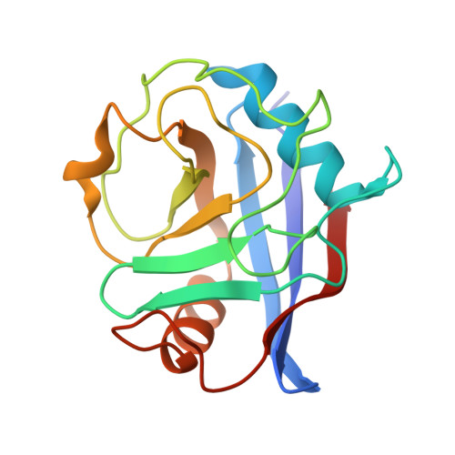
    9H0S, 9HTR

  • PubMed Abstract: 

    Cyclophilin D inhibitors that prevent opening of the mitochondrial permeability transition pore (MPTP) are potential treatments for a range of acute and chronic diseases, including acute pancreatitis. Here, we report that replacement of carbon with nitrogen in the pyrrolidine headgroup of a series of cyclophilin D inhibitors gives a dramatic enhancement in binding affinity (>40 fold), and prolyl isomerase inhibition (PPIase) activity (>200 fold), which is ascribed to a preorganization of the pyrazolidine amide headgroup. Protein-ligand X-ray crystal structures and NMR and molecular modeling demonstrate the importance of cis -amide geometry within the preorganized conformation, ensuring the ligand headgroup is anchored in the S1' binding pocket, leading to potent nM PPIase inhibition and binding. Pyrazolidines potently inhibit MPTP opening and prevent pancreatic toxin-induced cell necrosis in vitro . In vivo , 18f provided a significant improvement of acute pancreatitis biomarkers in the CER-AP mouse pancreatitis model, underlining the potential of this series.


  • Organizational Affiliation
    • Department of Molecular and Clinical Cancer Medicine, Liverpool Pancreatitis Research Group, University of Liverpool, Liverpool L69 7BE, U.K.

Macromolecule Content 

  • Total Structure Weight: 19.08 kDa 
  • Atom Count: 1,533 
  • Modeled Residue Count: 164 
  • Deposited Residue Count: 165 
  • Unique protein chains: 1

Macromolecules

Find similar proteins by:|  3D Structure
Entity ID: 1
MoleculeChains  Sequence LengthOrganismDetailsImage
Peptidyl-prolyl cis-trans isomerase F, mitochondrial165Homo sapiensMutation(s): 1 
Gene Names: PPIFCYP3
EC: 5.2.1.8
UniProt & NIH Common Fund Data Resources
Find proteins for P30405 (Homo sapiens)
Explore P30405 
Go to UniProtKB:  P30405
PHAROS:  P30405
GTEx:  ENSG00000108179 
Entity Groups
Sequence Clusters30% Identity50% Identity70% Identity90% Identity95% Identity100% Identity
UniProt GroupP30405
Sequence Annotations
Expand
Reference Sequence

Small Molecules

Ligands 6 Unique
IDChains Name / Formula / InChI Key2D Diagram3D Interactions
9CK
(Subject of Investigation/LOI)

Query on 9CK


Download:Ideal Coordinates CCD File
B [auth A]1-[(4-aminophenyl)methyl]-3-[2-[2-(2-bromophenyl)pyrazolidin-1-yl]-2-oxidanylidene-ethyl]urea
C19 H22 Br N5 O2
CDUGYVAEBXUUHW-UHFFFAOYSA-N
1PE

Query on 1PE


Download:Ideal Coordinates CCD File
H [auth A]PENTAETHYLENE GLYCOL
C10 H22 O6
JLFNLZLINWHATN-UHFFFAOYSA-N
PGE

Query on PGE


Download:Ideal Coordinates CCD File
I [auth A]TRIETHYLENE GLYCOL
C6 H14 O4
ZIBGPFATKBEMQZ-UHFFFAOYSA-N
PO4

Query on PO4


Download:Ideal Coordinates CCD File
J [auth A]PHOSPHATE ION
O4 P
NBIIXXVUZAFLBC-UHFFFAOYSA-K
EDO

Query on EDO


Download:Ideal Coordinates CCD File
C [auth A],
D [auth A],
E [auth A],
F [auth A],
G [auth A]
1,2-ETHANEDIOL
C2 H6 O2
LYCAIKOWRPUZTN-UHFFFAOYSA-N
NA

Query on NA


Download:Ideal Coordinates CCD File
K [auth A],
L [auth A],
M [auth A]
SODIUM ION
Na
FKNQFGJONOIPTF-UHFFFAOYSA-N

Experimental Data & Validation

Experimental Data

  • Method: X-RAY DIFFRACTION
  • Resolution: 1.45 Å
  • R-Value Free:  0.195 (Depositor), 0.198 (DCC) 
  • R-Value Work:  0.168 (Depositor), 0.172 (DCC) 
  • R-Value Observed: 0.170 (Depositor) 
Space Group: P 43 21 2
Unit Cell:
Length ( Å )Angle ( ˚ )
a = 57.45α = 90
b = 57.45β = 90
c = 114.91γ = 90
Software Package:
Software NamePurpose
PHENIXrefinement
iMOSFLMdata reduction
SCALAdata scaling
PHASERphasing

Structure Validation

View Full Validation Report


Ligand Structure Quality Assessment 


Entry History 

& Funding Information

Deposition Data


Funding OrganizationLocationGrant Number
Not fundedUnited Kingdom--

Revision History  (Full details and data files)

  • Version 1.0: 2025-11-05
    Type: Initial release
  • Version 1.1: 2025-12-17
    Changes: Database references