


Crystal Structure of Pyrophosphate-fructose 6-phosphate 1-phosphotransferase 1 (Pfk1) from Trichomonas vaginalis (AMP/alpha-D-Glucose-6-phosphate complex)
Liu, L., Lovell, S., Battaile, K.P.To be published.
Experimental Data Snapshot
Starting Model: experimental
View more details
Entity ID: 1 | |||||
|---|---|---|---|---|---|
| Molecule | Chains | Sequence Length | Organism | Details | Image |
| Pyrophosphate--fructose 6-phosphate 1-phosphotransferase 1 | 434 | Trichomonas vaginalis G3 | Mutation(s): 0 Gene Names: Pfk1, TVAG_430830 EC: 2.7.1.90 | 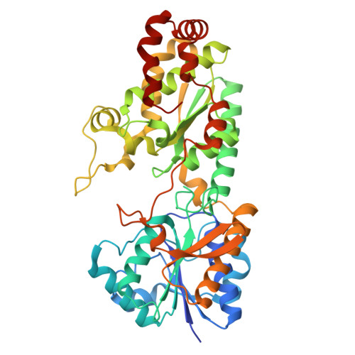 | |
UniProt | |||||
Entity Groups | |||||
| Sequence Clusters | 30% Identity50% Identity70% Identity90% Identity95% Identity100% Identity | ||||
| UniProt Group | O61068 | ||||
Sequence AnnotationsExpand | |||||
Reference Sequence | |||||
| Ligands 7 Unique | |||||
|---|---|---|---|---|---|
| ID | Chains | Name / Formula / InChI Key | 2D Diagram | 3D Interactions | |
| AMP (Subject of Investigation/LOI) Download:Ideal Coordinates CCD File | E [auth A], F [auth A], N [auth B], V [auth D] | ADENOSINE MONOPHOSPHATE C10 H14 N5 O7 P UDMBCSSLTHHNCD-KQYNXXCUSA-N |  | ||
| G6P (Subject of Investigation/LOI) Download:Ideal Coordinates CCD File | I [auth A], O [auth B] | 6-O-phosphono-alpha-D-glucopyranose C6 H13 O9 P NBSCHQHZLSJFNQ-DVKNGEFBSA-N |  | ||
| PPV (Subject of Investigation/LOI) Download:Ideal Coordinates CCD File | G [auth A] | PYROPHOSPHATE H4 O7 P2 XPPKVPWEQAFLFU-UHFFFAOYSA-N |  | ||
| PO4 Download:Ideal Coordinates CCD File | J [auth A] K [auth A] P [auth B] Q [auth B] R [auth B] | PHOSPHATE ION O4 P NBIIXXVUZAFLBC-UHFFFAOYSA-K |  | ||
| GOL Download:Ideal Coordinates CCD File | H [auth A] | GLYCEROL C3 H8 O3 PEDCQBHIVMGVHV-UHFFFAOYSA-N |  | ||
| CL Download:Ideal Coordinates CCD File | L [auth A] | CHLORIDE ION Cl VEXZGXHMUGYJMC-UHFFFAOYSA-M |  | ||
| MG Download:Ideal Coordinates CCD File | M [auth A] | MAGNESIUM ION Mg JLVVSXFLKOJNIY-UHFFFAOYSA-N |  | ||
| Length ( Å ) | Angle ( ˚ ) |
|---|---|
| a = 86.909 | α = 90 |
| b = 86.909 | β = 90 |
| c = 473.809 | γ = 90 |
| Software Name | Purpose |
|---|---|
| PHENIX | refinement |
| Aimless | data scaling |
| XDS | data reduction |
| PHASER | phasing |
| Funding Organization | Location | Grant Number |
|---|---|---|
| National Institutes of Health/National Institute Of Allergy and Infectious Diseases (NIH/NIAID) | United States | 75N93022C00036 |
| National Institutes of Health/Office of the Director | United States | S10OD030394 |


