


Structure of SARM1 TIR domain bound to G6831
Wallweber, H.A., Sudhamsu, J.To be published.
Experimental Data Snapshot
Starting Model: experimental
View more details
Entity ID: 1 | |||||
|---|---|---|---|---|---|
| Molecule | Chains | Sequence Length | Organism | Details | Image |
| NAD(+) hydrolase SARM1 | 143 | Homo sapiens | Mutation(s): 0 Gene Names: SARM1, KIAA0524, SAMD2, SARM EC: 3.2.2.6 (PDB Primary Data), 3.2.2 (PDB Primary Data) | 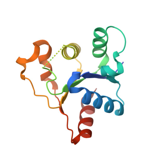 | |
UniProt & NIH Common Fund Data Resources | |||||
PHAROS: Q6SZW1 GTEx: ENSG00000004139 | |||||
Entity Groups | |||||
| Sequence Clusters | 30% Identity50% Identity70% Identity90% Identity95% Identity100% Identity | ||||
| UniProt Group | Q6SZW1 | ||||
Sequence AnnotationsExpand | |||||
Reference Sequence | |||||
| Ligands 1 Unique | |||||
|---|---|---|---|---|---|
| ID | Chains | Name / Formula / InChI Key | 2D Diagram | 3D Interactions | |
| A1BU7 (Subject of Investigation/LOI) Download:Ideal Coordinates CCD File | C [auth A], D [auth B] | [[(2~{R},3~{S},4~{R},5~{R})-5-(6-aminopurin-9-yl)-3,4-bis(oxidanyl)oxolan-2-yl]methoxy-oxidanyl-phosphoryl] [(2~{R},3~{S},4~{R},5~{R})-5-[4-[3-[(4~{S})-4-(4-chloranyl-3-fluoranyl-phenyl)-2-oxidanylidene-piperidin-1-yl]-1-bicyclo[1.1.1]pentanyl]pyridin-1-yl]-3,4-bis(oxidanyl)oxolan-2-yl]methyl hydrogen phosphate C36 H42 Cl F N7 O14 P2 MOFVGPYVZJGLNR-PXGPZXLQSA-O |  | ||
| Length ( Å ) | Angle ( ˚ ) |
|---|---|
| a = 33.115 | α = 90 |
| b = 85.818 | β = 90 |
| c = 116.957 | γ = 90 |
| Software Name | Purpose |
|---|---|
| PHENIX | refinement |
| XDS | data scaling |
| autoPROC | data reduction |
| PHENIX | phasing |
| Funding Organization | Location | Grant Number |
|---|---|---|
| F. Hoffmann-La Roche LTD | Switzerland | -- |


