Download Mendeley
Antibody CDR-H3 loop flexibility: Insights from X-ray crystallography, structural bioinformatics, and the limits of current deep learning methods.
Barozet, A., Mathieu, M., Papin, D., Cameron, B., Dabdoubi, T., Severac, A., Ferrari, P., Simeon, T., Bianciotto, M., Cortes, J.(2026) J Struct Biol 218: 108368-108368
- PubMed: 42731718 Search on PubMed
- DOI: https://doi.org/10.1016/j.jsb.2026.108368
- Primary Citation Related Structures:
9TVR, 9TVZ, 9TW1, 9TW7 - PubMed Abstract:
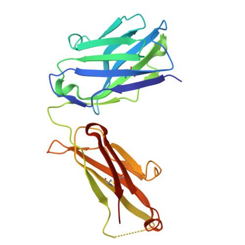
Complementarity Determining Regions (CDRs) in antibodies, and in particular the CDR-H3 loop, often display conformational plasticity that is essential for their function. Due to this flexibility, the structural investigation of antibody-antigen binding cannot exclusively rely on experimental techniques that only provide snapshots of unbound and bound states, such as X-ray crystallography. Moreover, X-ray structures can be biased due to experimental conditions and crystal packing. In this context, computational techniques, and especially conformational sampling methods, are an essential complement to experiments. This work illustrates the interest of such a coupling of methods on the structural investigation of an anti-FGFR4 (Fibroblast growth factor receptor 4) antibody. X-ray crystallography experiments revealed a very significant conformational change of the CDR-H3 loop between unbound and bound states. Structural bioinformatics methods were then applied to provide a more global picture of the conformational space of this loop, and to confirm that the observed conformations were not the result of experimental artifacts. The experimental unbound conformation was reliably predicted, and the loop conformation observed in the bound state was also predicted to be a probable conformation in the absence of the antigen. The possible existence of a third low-energy conformation, for which there is currently no experimental evidence, was substantiated by molecular simulations. We also applied recent methods based on deep learning techniques to evaluate their ability to predict conformations of the H3 loop. The results show that while these methods are very effective at predicting the structure of rigid/stable regions of proteins, they still have difficulties in accurately representing regions with more variable structure, such as this loop. Overall, this work shows that the structural study of flexible proteins remains an open field of research, and that the synergistic coupling of experimental and computational methods is essential in this context.
- Université de Toulouse, CNRS, LAAS, Toulouse, France; Sanofi Recherche & Développement, Integrated Drug Discovery, Molecular Design Sciences, 13 quai Jules Guesde, BP 14, 94403 Vitry-sur-Seine Cedex, France. Electronic address: abarozet@gmail.com.
Organizational Affiliation: