Download Mendeley
Retroviruses use different IP 6 binding mechanisms to alter the properties of their capsids.
Klarhof, J.O., Mallery, D.L., Stacey, J.C.V., Torre, D., Rumlova, M., Ruml, T., Briggs, J.A.G., James, L.C.(2026) Nat Commun 17
- PubMed: 42764334 Search on PubMedSearch on PubMed Central
- DOI: https://doi.org/10.1038/s41467-026-76510-7
- Primary Citation Related Structures:
9TX4, 9TX5, 9TX6, 9TX7, 9TX8, 9TX9, 9TXA, 9TXB - PubMed Abstract:
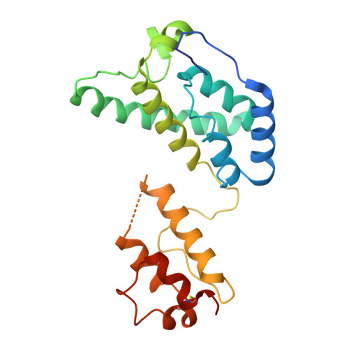
HIV-1 uses the metabolite inositol hexakisphosphate (IP 6 ) as a host factor to assemble its capsid, but whether this strategy is unique to lentiviruses or represents a common feature of retroviral capsids remains unclear. Here we show that IP 6 binding is conserved across diverse retroviruses but occurs through distinct capsid sites and mechanisms, and influences viral behaviour. In contrast to HIV-1, the beta-retrovirus Mason-Pfizer Monkey Virus (MPMV) and the gamma-retrovirus Murine Leukaemia Virus (MLV) bind IP 6 at the threefold lattice interface between capsomers rather than within capsomer pores. Cryo-EM structures of core-like particles reveal that two lysine residues from each capsomer coordinate IP 6 between either two discrete three-lysine rings (MPMV) or a single heterogeneous six-lysine ring (MLV). MPMV and MLV are largely insensitive to IP 6 availability in producer cells, but this binding mode renders them highly dependent on IP6 in target cells - the opposite of the dependency pattern of HIV-1. The way in which retroviruses use IP 6 to build their capsids alters their dependence on the metabolite at different stages of the replicative cycle and in key capsid behaviours, such as assembly and stability.
- MRC Laboratory of Molecular Biology, Cambridge, UK.
Organizational Affiliation: