9VNS | pdb_00009vns

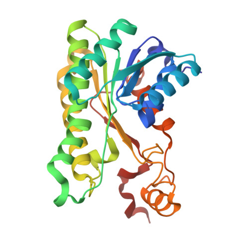
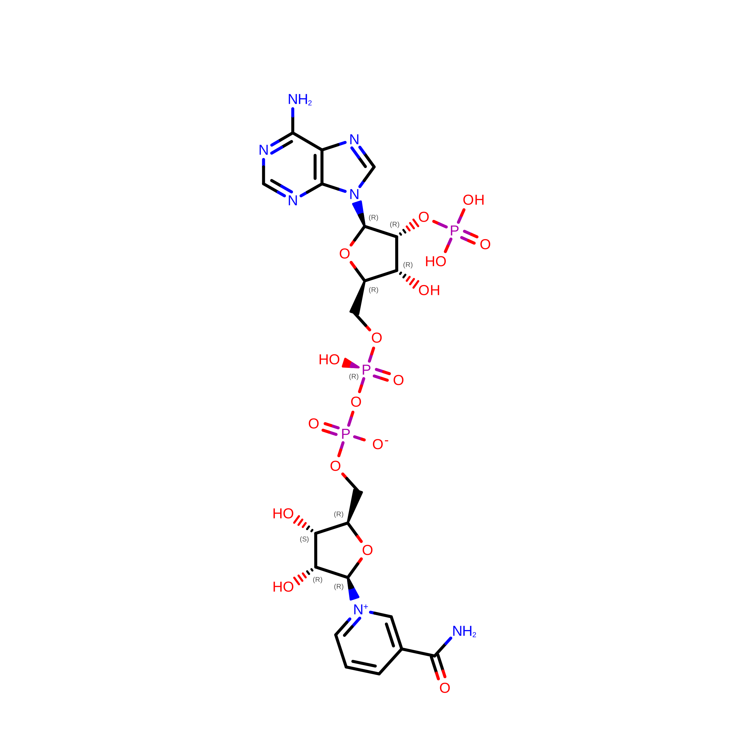
NADPH and 1-benzyl-4-methylpiperidin-3-one complex structure of glucose dehydrogenase M6 from Bacillus megaterium


Experimental Data Snapshot

  • Method: X-RAY DIFFRACTION
  • Resolution: 2.09 Å
  • R-Value Free: 
    0.267 (Depositor), 0.266 (DCC) 
  • R-Value Work: 
    0.214 (Depositor), 0.214 (DCC) 
  • R-Value Observed: 
    0.216 (Depositor) 

Starting Model: experimental
View more details

wwPDB Validation 3D Report Full Report

Validation slider image for 9VNS

Ligand Structure Quality Assessment 


This is version 1.0 of the entry. See complete history. 

Literature

Crystal structure of glucose dehydrogenase from Bacillus megaterium IWG3 at 1.7 A resolution.

Yamamoto, K., Kurisu, G., Kusunoki, M., Tabata, S., Urabe, I., Osaki, S.

(2001) J Biochem 129: 303-312

  • DOI: https://doi.org/10.1093/oxfordjournals.jbchem.a002858
  • Primary Citation Related Structures: 
    1GCO, 9VNS, 9VP1

  • PubMed Abstract: 

    The crystal structure of glucose dehydrogenase (GlcDH) from Bacillus megaterium IWG3 has been determined to an R-factor of 17.9% at 1.7 A resolution. The enzyme consists of four identical subunits, which are similar to those of other short-chain reductases/dehydrogenases (SDRs) in their overall folding and subunit architecture, although cofactor binding sites and subunit interactions differ. Whereas a pair of basic residues is well conserved among NADP(+)-preferring SDRs, only Arg39 was found around the adenine ribose moiety of GlcDH. This suggests that one basic amino acid is enough to determine the coenzyme specificity. The four subunits are interrelated by three mutually perpendicular diad axes (P, Q, and R). While subunit interactions through the P-axis for GlcDH are not so different from those of the other SDRs, those through the Q-axis differ significantly. GlcDH was found to have weaker hydrophobic interactions in the Q-interface. Moreover, GlcDH lacks the salt bridge that stabilizes the subunit interaction in the Q-interface in the other SDRs. Hydrogen bonds between Q-axis related subunits are also less common than in the other SDRs. The GlcDH tetramer dissociates into inactive monomers at pH 9.0, which can be attributed mainly to the weakness of the Q-axis interface.


  • Organizational Affiliation: 
    • Department of Chemistry, Nara Medical University, Shijo, Kashihara, Nara 634-8521, Japan. kama@naramed-u.ac.jp

Macromolecule Content 

  • Total Structure Weight: 117.08 kDa 
  • Atom Count: 8,258 
  • Modeled Residue Count: 1,040 
  • Deposited Residue Count: 1,052 
  • Unique protein chains: 1

Macromolecules

Find similar proteins by:|  3D Structure
Entity ID: 1
MoleculeChains  Sequence LengthOrganismDetailsImage
Glucose 1-dehydrogenase
A, B, C, D
263Priestia megateriumMutation(s): 0 
EC: 1.1.1.47
UniProt
Find proteins for P40288 (Priestia megaterium)
Explore P40288 
Go to UniProtKB:  P40288
Entity Groups
Sequence Clusters30% Identity50% Identity70% Identity90% Identity95% Identity100% Identity
UniProt GroupP40288
Sequence Annotations
Expand
Reference Sequence

Experimental Data & Validation

Experimental Data

  • Method: X-RAY DIFFRACTION
  • Resolution: 2.09 Å
  • R-Value Free:  0.267 (Depositor), 0.266 (DCC) 
  • R-Value Work:  0.214 (Depositor), 0.214 (DCC) 
  • R-Value Observed: 0.216 (Depositor) 
Space Group: P 32
Unit Cell:
Length ( Å )Angle ( ˚ )
a = 71.18α = 90
b = 71.18β = 90
c = 175.46γ = 120
Software Package:
Software NamePurpose
PHENIXrefinement
PHENIXrefinement
xia2data reduction
xia2data scaling
REFMACphasing

Structure Validation

View Full Validation Report


Ligand Structure Quality Assessment 


Entry History 

& Funding Information

Deposition Data


Funding OrganizationLocationGrant Number
National Natural Science Foundation of China (NSFC)China--

Revision History  (Full details and data files)

  • Version 1.0: 2026-10-07
    Type: Initial release