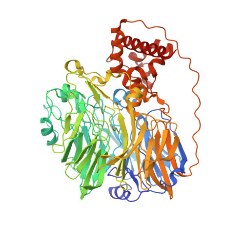
1KV9 | pdb_00001kv9

Structure at 1.9 A Resolution of a Quinohemoprotein Alcohol Dehydrogenase from Pseudomonas putida HK5


Experimental Data Snapshot

  • Method: X-RAY DIFFRACTION
  • Resolution: 1.90 Å
  • R-Value Free: 
    0.189 (Depositor), 0.180 (DCC) 
  • R-Value Work: 
    0.154 (Depositor), 0.150 (DCC) 

Starting Model: experimental
View more details

wwPDB Validation   3D Report Full Report


Ligand Structure Quality Assessment 

Created with Raphaël 2.3.0Worse 01 BetterLigand structure goodness of fit to experimental dataBest fitted HECClick on this verticalbar to view detailsBest fitted PQQClick on this verticalbar to view details

This is version 2.2 of the entry. See complete history


Literature

Structure at 1.9 A resolution of a quinohemoprotein alcohol dehydrogenase from Pseudomonas putida HK5.

Chen, Z.W.Matsushita, K.Yamashita, T.Fujii, T.A.Toyama, H.Adachi, O.Bellamy, H.D.Mathews, F.S.

(2002) Structure 10: 837-849

  • DOI: https://doi.org/10.1016/s0969-2126(02)00774-8
  • Primary Citation of Related Structures:  
    1KV9

  • PubMed Abstract: 

    The type II quinohemoprotein alcohol dehydrogenase of Pseudomonas putida is a periplasmic enzyme that oxidizes substrate alcohols to the aldehyde and transfers electrons first to pyrroloquinoline quinone (PQQ) and then to an internal heme group. The 1.9 A resolution crystal structure reveals that the enzyme contains a large N-terminal eight-stranded beta propeller domain (approximately 60 kDa) similar to methanol dehydrogenase and a small C-terminal c-type cytochrome domain (approximately 10 kDa) similar to the cytochrome subunit of p-cresol methylhydoxylase. The PQQ is bound near the axis of the propeller domain about 14 A from the heme. A molecule of acetone, the product of the oxidation of isopropanol present during crystallization, appears to be bound in the active site cavity.


  • Organizational Affiliation

    Department of Biochemistry and Molecular Biophysics, Washington University School of Medicine, St. Louis, MO 63110, USA.


Macromolecules
Find similar proteins by:  (by identity cutoff)  |  3D Structure
Entity ID: 1
MoleculeChains Sequence LengthOrganismDetailsImage
TYPE II QUINOHEMOPROTEIN ALCOHOL DEHYDROGENASE668Pseudomonas putidaMutation(s): 0 
EC: 1.1.99 (PDB Primary Data), 1.1.9.1 (UniProt)
Membrane Entity: Yes 
UniProt
Find proteins for Q8GR64 (Pseudomonas putida)
Explore Q8GR64 
Go to UniProtKB:  Q8GR64
Entity Groups  
Sequence Clusters30% Identity50% Identity70% Identity90% Identity95% Identity100% Identity
UniProt GroupQ8GR64
Sequence Annotations
Expand
  • Reference Sequence
Small Molecules
Ligands 6 Unique
IDChains Name / Formula / InChI Key2D Diagram3D Interactions
HEC
Query on HEC

Download Ideal Coordinates CCD File 
D [auth A]HEME C
C34 H34 Fe N4 O4
HXQIYSLZKNYNMH-LJNAALQVSA-N
PQQ
Query on PQQ

Download Ideal Coordinates CCD File 
C [auth A]PYRROLOQUINOLINE QUINONE
C14 H6 N2 O8
MMXZSJMASHPLLR-UHFFFAOYSA-N
EPE
Query on EPE

Download Ideal Coordinates CCD File 
E [auth A]4-(2-HYDROXYETHYL)-1-PIPERAZINE ETHANESULFONIC ACID
C8 H18 N2 O4 S
JKMHFZQWWAIEOD-UHFFFAOYSA-N
GOL
Query on GOL

Download Ideal Coordinates CCD File 
G [auth A]GLYCEROL
C3 H8 O3
PEDCQBHIVMGVHV-UHFFFAOYSA-N
ACN
Query on ACN

Download Ideal Coordinates CCD File 
F [auth A]ACETONE
C3 H6 O
CSCPPACGZOOCGX-UHFFFAOYSA-N
CA
Query on CA

Download Ideal Coordinates CCD File 
B [auth A]CALCIUM ION
Ca
BHPQYMZQTOCNFJ-UHFFFAOYSA-N
Experimental Data & Validation

Experimental Data

  • Method: X-RAY DIFFRACTION
  • Resolution: 1.90 Å
  • R-Value Free:  0.189 (Depositor), 0.180 (DCC) 
  • R-Value Work:  0.154 (Depositor), 0.150 (DCC) 
Space Group: P 1
Unit Cell:
Length ( Å )Angle ( ˚ )
a = 54.807α = 89.65
b = 57.437β = 69.34
c = 67.523γ = 68.39
Software Package:
Software NamePurpose
DENZOdata reduction
SCALEPACKdata scaling
AMoREphasing
CNSrefinement
SHARPphasing

Structure Validation

View Full Validation Report


Ligand Structure Quality Assessment 

Created with Raphaël 2.3.0Worse 01 BetterLigand structure goodness of fit to experimental dataBest fitted HECClick on this verticalbar to view detailsBest fitted PQQClick on this verticalbar to view details

Entry History 

Deposition Data

Revision History  (Full details and data files)

  • Version 1.0: 2002-07-10
    Type: Initial release
  • Version 1.1: 2008-04-27
    Changes: Version format compliance
  • Version 1.2: 2011-07-13
    Changes: Non-polymer description, Version format compliance
  • Version 2.0: 2021-03-03
    Changes: Advisory, Atomic model, Data collection, Derived calculations, Non-polymer description, Structure summary
  • Version 2.1: 2023-08-16
    Changes: Data collection, Database references, Refinement description
  • Version 2.2: 2024-11-06
    Changes: Structure summary