1P5R | pdb_00001p5r

Formyl-CoA Transferase in complex with Coenzyme A


Experimental Data Snapshot

  • Method: X-RAY DIFFRACTION
  • Resolution: 2.50 Å
  • R-Value Free: 
    0.236 (Depositor), 0.236 (DCC) 
  • R-Value Work: 
    0.194 (Depositor), 0.194 (DCC) 
  • R-Value Observed: 
    0.194 (Depositor) 

Starting Model: experimental
View more details

wwPDB Validation 3D Report Full Report

Validation slider image for 1P5R

Ligand Structure Quality Assessment 


This is version 1.3 of the entry. See complete history

Literature

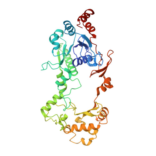
Formyl-CoA Transferase encloses the CoA binding site at the interface of an interlocked dimer

Ricagno, S.Jonsson, S.Richards, N.Lindqvist, Y.

(2003) EMBO J 22: 3210-3219

  • DOI: https://doi.org/10.1093/emboj/cdg333
  • Primary Citation Related Structures: 
    1P5H, 1P5R

  • PubMed Abstract: 

    Formyl-CoA transferase catalyses transfer of CoA from formate to oxalate in the first step of oxalate degradation by Oxalobacter formigenes, a bacterium present in the intestinal flora which is implicated in oxalate catabolism in mammals. Formyl-CoA transferase is a member of a family of CoA-transferases for which no structural information is available. We now report the three-dimensional structure of O.formigenes formyl-CoA transferase, which reveals a novel fold and a very striking assembly of the homodimer. The subunit is composed of a large and a small domain where residues from both the N- and C-termini of the subunit are part of the large domain. The linkers between the domains give the subunit a circular shape with a hole in the middle. The enzyme monomers are tightly interacting and are interlocked. This fold requires drastic rearrangement of approximately 75 residues at the C-terminus for formation of the dimer. The structure of a complex of formyl-CoA transferase with CoA is also reported and sets the scene for a mechanistic understanding of enzymes of this family of CoA-transferases.


  • Organizational Affiliation
    • Department of Medical Biochemistry and Biophysics, Karolinska Institutet, S-17177 Stockholm, Sweden.

Macromolecule Content 

  • Total Structure Weight: 96.3 kDa 
  • Atom Count: 6,982 
  • Modeled Residue Count: 854 
  • Deposited Residue Count: 856 
  • Unique protein chains: 1

Macromolecules

Find similar proteins by:|  3D Structure
Entity ID: 1
MoleculeChains  Sequence LengthOrganismDetailsImage
Formyl-coenzyme A transferase
A, B
428Oxalobacter formigenesMutation(s): 0 
Gene Names: FRC
EC: 2.8.3 (PDB Primary Data), 2.8.3.16 (UniProt)
UniProt
Find proteins for O06644 (Oxalobacter formigenes)
Explore O06644 
Go to UniProtKB:  O06644
Entity Groups
Sequence Clusters30% Identity50% Identity70% Identity90% Identity95% Identity100% Identity
UniProt GroupO06644
Sequence Annotations
Expand
Reference Sequence

Experimental Data & Validation

Experimental Data

  • Method: X-RAY DIFFRACTION
  • Resolution: 2.50 Å
  • R-Value Free:  0.236 (Depositor), 0.236 (DCC) 
  • R-Value Work:  0.194 (Depositor), 0.194 (DCC) 
  • R-Value Observed: 0.194 (Depositor) 
Space Group: I 4
Unit Cell:
Length ( Å )Angle ( ˚ )
a = 151.4α = 90
b = 151.4β = 90
c = 99.5γ = 90
Software Package:
Software NamePurpose
CNSrefinement
MOSFLMdata reduction
CCP4data scaling
CNSphasing

Structure Validation

View Full Validation Report


Ligand Structure Quality Assessment 


Entry History 

Deposition Data

Revision History  (Full details and data files)

  • Version 1.0: 2003-07-29
    Type: Initial release
  • Version 1.1: 2008-04-29
    Changes: Version format compliance
  • Version 1.2: 2011-07-13
    Changes: Version format compliance
  • Version 1.3: 2023-08-16
    Changes: Data collection, Database references, Derived calculations, Refinement description