1S22

Absolute Stereochemistry of Ulapualide A


Experimental Data Snapshot

  • Method: X-RAY DIFFRACTION
  • Resolution: 1.60 Å
  • R-Value Free: 
    0.179 (Depositor), 0.190 (DCC) 
  • R-Value Work: 
    0.152 (Depositor), 0.170 (DCC) 
  • R-Value Observed: 
    0.153 (Depositor) 

Starting Model: experimental
View more details

wwPDB Validation   3D Report Full Report


Ligand Structure Quality Assessment 

Created with Raphaël 2.3.0Worse 01 BetterLigand structure goodness of fit to experimental dataBest fitted ULAClick on this verticalbar to view detailsBest fitted ATPClick on this verticalbar to view details

This is version 1.3 of the entry. See complete history


Literature

Absolute stereochemistry of ulapualide A

Allingham, J.S.Tanaka, J.Marriott, G.Rayment, I.

(2004) Org Lett 6: 597-599

  • DOI: https://doi.org/10.1021/ol036458y
  • Primary Citation of Related Structures:  
    1S22

  • PubMed Abstract: 

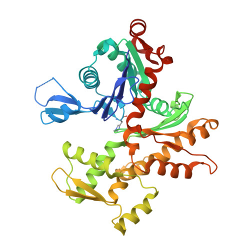
    [structure: see text] The structure of ulapualide A (1) has been solved by X-ray crystallography in a complex with G-actin. The stereochemical configuration was assigned as 3S,9S,22S,23R,24S,26S,27S,31R,32R,33R.


  • Organizational Affiliation

    Department of Biochemistry, University of Wisconsin-Madison, Madison, Wisconsin 53706, USA.


Macromolecules
Find similar proteins by:  (by identity cutoff)  |  3D Structure
Entity ID: 1
MoleculeChains Sequence LengthOrganismDetailsImage
Actin375Oryctolagus cuniculusMutation(s): 1 
EC: 3.6.4
UniProt
Find proteins for P68135 (Oryctolagus cuniculus)
Explore P68135 
Go to UniProtKB:  P68135
Entity Groups  
Sequence Clusters30% Identity50% Identity70% Identity90% Identity95% Identity100% Identity
UniProt GroupP68135
Sequence Annotations
Expand
  • Reference Sequence
Small Molecules
Ligands 4 Unique
IDChains Name / Formula / InChI Key2D Diagram3D Interactions
ULA
Query on ULA

Download Ideal Coordinates CCD File 
D [auth A]ULAPUALIDE A
C46 H66 N4 O13
DIOFXPZEAVIPDB-GSMFZUCRSA-N
ATP
Query on ATP

Download Ideal Coordinates CCD File 
C [auth A]ADENOSINE-5'-TRIPHOSPHATE
C10 H16 N5 O13 P3
ZKHQWZAMYRWXGA-KQYNXXCUSA-N
EDO
Query on EDO

Download Ideal Coordinates CCD File 
E [auth A],
F [auth A]
1,2-ETHANEDIOL
C2 H6 O2
LYCAIKOWRPUZTN-UHFFFAOYSA-N
CA
Query on CA

Download Ideal Coordinates CCD File 
B [auth A]CALCIUM ION
Ca
BHPQYMZQTOCNFJ-UHFFFAOYSA-N
Modified Residues  1 Unique
IDChains TypeFormula2D DiagramParent
HIC
Query on HIC
A
L-PEPTIDE LINKINGC7 H11 N3 O2HIS
Experimental Data & Validation

Experimental Data

  • Method: X-RAY DIFFRACTION
  • Resolution: 1.60 Å
  • R-Value Free:  0.179 (Depositor), 0.190 (DCC) 
  • R-Value Work:  0.152 (Depositor), 0.170 (DCC) 
  • R-Value Observed: 0.153 (Depositor) 
Space Group: P 1 21 1
Unit Cell:
Length ( Å )Angle ( ˚ )
a = 40.8α = 90
b = 75.2β = 100.2
c = 67.5γ = 90
Software Package:
Software NamePurpose
HKL-2000data collection
SCALEPACKdata scaling
MOLREPphasing
REFMACrefinement
HKL-2000data reduction

Structure Validation

View Full Validation Report


Ligand Structure Quality Assessment 

Created with Raphaël 2.3.0Worse 01 BetterLigand structure goodness of fit to experimental dataBest fitted ULAClick on this verticalbar to view detailsBest fitted ATPClick on this verticalbar to view details

Entry History 

Deposition Data

Revision History  (Full details and data files)

  • Version 1.0: 2004-02-17
    Type: Initial release
  • Version 1.1: 2008-04-29
    Changes: Version format compliance
  • Version 1.2: 2011-07-13
    Changes: Version format compliance
  • Version 1.3: 2023-08-23
    Changes: Data collection, Database references, Derived calculations, Refinement description