2C54

gdp-mannose-3', 5' -epimerase (arabidopsis thaliana),k178r, with gdp-beta-l-gulose and gdp-4-keto-beta-l-gulose bound in active site.


Experimental Data Snapshot

  • Method: X-RAY DIFFRACTION
  • Resolution: 1.50 Å
  • R-Value Free: 
    0.167 (Depositor), 0.170 (DCC) 
  • R-Value Work: 
    0.118 (Depositor), 0.130 (DCC) 
  • R-Value Observed: 
    0.121 (Depositor) 

wwPDB Validation   3D Report Full Report


Ligand Structure Quality Assessment 

Created with Raphaël 2.3.0Worse 01 BetterLigand structure goodness of fit to experimental dataBest fitted NADClick on this verticalbar to view detailsBest fitted GKEClick on this verticalbar to view detailsBest fitted GKDClick on this verticalbar to view details

This is version 1.6 of the entry. See complete history


Literature

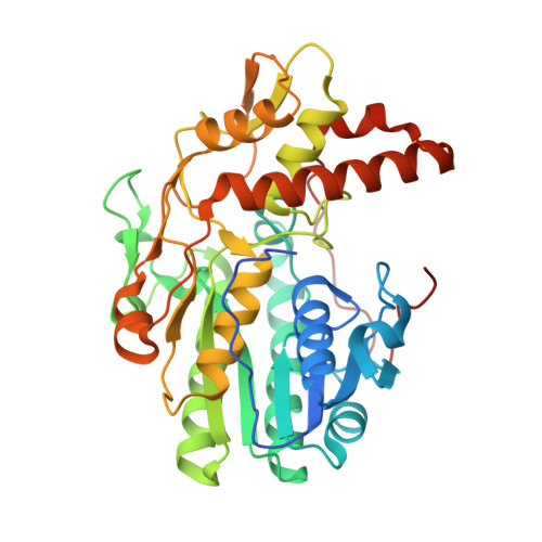
Structure and function of GDP-mannose-3',5'-epimerase: an enzyme which performs three chemical reactions at the same active site.

Major, L.L.Wolucka, B.A.Naismith, J.H.

(2005) J Am Chem Soc 127: 18309-18320

  • DOI: https://doi.org/10.1021/ja056490i
  • Primary Citation of Related Structures:  
    2C54, 2C59, 2C5A, 2C5E

  • PubMed Abstract: 

    GDP-mannose-3',5'-epimerase (GME) from Arabidopsis thaliana catalyzes the epimerization of both the 3' and 5' positions of GDP-alpha-D-mannose to yield GDP-beta-L-galactose. Production of the C5' epimer of GDP-alpha-D-mannose, GDP-beta-L-gulose, has also been reported. The reaction occurs as part of vitamin C biosynthesis in plants. We have determined structures of complexes of GME with GDP-alpha-D-mannose, GDP-beta-L-galactose, and a mixture of GDP-beta-L-gulose with GDP-beta-L-4-keto-gulose to resolutions varying from 2.0 to 1.4 A. The enzyme has the classical extended short-chain dehydratase/reductase (SDR) fold. We have confirmed that GME establishes an equilibrium between two products, GDP-beta-L-galactose and GDP-beta-L-gulose. The reaction proceeds by C4' oxidation of GDP-alpha-D-mannose followed by epimerization of the C5' position to give GDP-beta-L-4-keto-gulose. This intermediate is either reduced to give GDP-beta-L-gulose or the C3' position is epimerized to give GDP-beta-L-4-keto-galactose, then C4' is reduced to GDP-beta-L-galactose. The combination of oxidation, epimerization, and reduction in a single active site is unusual. Structural analysis coupled to site-directed mutagenesis suggests C145 and K217 as the acid/base pair responsible for both epimerizations. On the basis of the structure of the GDP-beta-L-gulose/GDP-beta-L-4-keto-gulose co-complex, we predict that a ring flip occurs during the first epimerization and that a boat intermediate is likely for the second epimerization. Comparison of GME with other SDR enzymes known to abstract a protein alpha to the keto function of a carbohydrate identifies key common features.


  • Organizational Affiliation

    Centre for Biomolecular Sciences, University of St. Andrews, North Haugh, St. Andrews, Fife, Scotland KY16 9ST, United Kingdom.


Macromolecules
Find similar proteins by:  (by identity cutoff)  |  3D Structure
Entity ID: 1
MoleculeChains Sequence LengthOrganismDetailsImage
GDP-MANNOSE-3', 5'-EPIMERASE
A, B
379Arabidopsis thalianaMutation(s): 1 
EC: 5.1.3.18
UniProt
Find proteins for Q93VR3 (Arabidopsis thaliana)
Explore Q93VR3 
Go to UniProtKB:  Q93VR3
Entity Groups  
Sequence Clusters30% Identity50% Identity70% Identity90% Identity95% Identity100% Identity
UniProt GroupQ93VR3
Sequence Annotations
Expand
  • Reference Sequence
Small Molecules
Ligands 5 Unique
IDChains Name / Formula / InChI Key2D Diagram3D Interactions
NAD
Query on NAD

Download Ideal Coordinates CCD File 
E [auth A],
R [auth B]
NICOTINAMIDE-ADENINE-DINUCLEOTIDE
C21 H27 N7 O14 P2
BAWFJGJZGIEFAR-NNYOXOHSSA-N
GKE
Query on GKE

Download Ideal Coordinates CCD File 
C [auth A],
P [auth B]
GUANOSINE 5'-DIPHOSPHATE-BETA-L-GULOSE
C16 H25 N5 O16 P2
MVMSCBBUIHUTGJ-HVMPVDAASA-N
GKD
Query on GKD

Download Ideal Coordinates CCD File 
D [auth A],
Q [auth B]
GUANOSINE 5'-DIPHOSPHATE-4-KETO-BETA-L-GULOSE
C16 H23 N5 O16 P2
QZZKIVADHWVJFF-UFFCYROPSA-N
EPE
Query on EPE

Download Ideal Coordinates CCD File 
O [auth A]4-(2-HYDROXYETHYL)-1-PIPERAZINE ETHANESULFONIC ACID
C8 H18 N2 O4 S
JKMHFZQWWAIEOD-UHFFFAOYSA-N
FMT
Query on FMT

Download Ideal Coordinates CCD File 
AA [auth B]
F [auth A]
G [auth A]
H [auth A]
I [auth A]
FORMIC ACID
C H2 O2
BDAGIHXWWSANSR-UHFFFAOYSA-N
Experimental Data & Validation

Experimental Data

  • Method: X-RAY DIFFRACTION
  • Resolution: 1.50 Å
  • R-Value Free:  0.167 (Depositor), 0.170 (DCC) 
  • R-Value Work:  0.118 (Depositor), 0.130 (DCC) 
  • R-Value Observed: 0.121 (Depositor) 
Space Group: P 1 21 1
Unit Cell:
Length ( Å )Angle ( ˚ )
a = 62.278α = 90
b = 82.457β = 98.77
c = 65.875γ = 90
Software Package:
Software NamePurpose
REFMACrefinement
MOSFLMdata reduction
SCALAdata scaling
PHASERphasing

Structure Validation

View Full Validation Report


Ligand Structure Quality Assessment 

Created with Raphaël 2.3.0Worse 01 BetterLigand structure goodness of fit to experimental dataBest fitted NADClick on this verticalbar to view detailsBest fitted GKEClick on this verticalbar to view detailsBest fitted GKDClick on this verticalbar to view details

Entry History 

Deposition Data

Revision History  (Full details and data files)

  • Version 1.0: 2005-11-14
    Type: Initial release
  • Version 1.1: 2011-05-08
    Changes: Version format compliance
  • Version 1.2: 2011-07-13
    Changes: Version format compliance
  • Version 1.3: 2018-03-07
    Changes: Source and taxonomy
  • Version 1.4: 2018-12-19
    Changes: Data collection, Database references, Derived calculations
  • Version 1.5: 2019-01-30
    Changes: Data collection, Experimental preparation
  • Version 1.6: 2024-05-08
    Changes: Data collection, Database references, Other