2CW8 | pdb_00002cw8

Crystal structure of intein homing endonuclease II


Experimental Data Snapshot

  • Method: X-RAY DIFFRACTION
  • Resolution: 2.50 Å
  • R-Value Free: 
    0.252 (Depositor), 0.260 (DCC) 
  • R-Value Work: 
    0.217 (Depositor), 0.226 (DCC) 
  • R-Value Observed: 
    0.217 (Depositor) 

wwPDB Validation 3D Report Full Report

Validation slider image for 2CW8

This is version 1.3 of the entry. See complete history

Literature

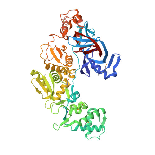
Crystal structure of intein homing endonuclease II encoded in DNA polymerase gene from hyperthermophilic archaeon Thermococcus kodakaraensis strain KOD1

Matsumura, H.Takahashi, H.Inoue, T.Yamamoto, T.Hashimoto, H.Nishioka, M.Fujiwara, S.Takagi, M.Imanaka, T.Kai, Y.

(2006) Proteins 63: 711-715

Macromolecule Content 

  • Total Structure Weight: 63.84 kDa 
  • Atom Count: 4,735 
  • Modeled Residue Count: 537 
  • Deposited Residue Count: 537 
  • Unique protein chains: 1

Macromolecules

Find similar proteins by:|  3D Structure
Entity ID: 1
MoleculeChains  Sequence LengthOrganismDetailsImage
Endonuclease PI-PkoII537Thermococcus kodakarensis KOD1Mutation(s): 9 
EC: 3.1 (PDB Primary Data), 2.7.7.7 (UniProt)
UniProt
Find proteins for P77933 (Thermococcus kodakarensis (strain ATCC BAA-918 / JCM 12380 / KOD1))
Explore P77933 
Go to UniProtKB:  P77933
Entity Groups
Sequence Clusters30% Identity50% Identity70% Identity90% Identity95% Identity100% Identity
UniProt GroupP77933
Sequence Annotations
Expand
Reference Sequence

Small Molecules

Ligands 2 Unique
IDChains Name / Formula / InChI Key2D Diagram3D Interactions
SO4

Query on SO4


Download:Ideal Coordinates CCD File
B [auth A]
C [auth A]
D [auth A]
E [auth A]
F [auth A]
B [auth A],
C [auth A],
D [auth A],
E [auth A],
F [auth A],
G [auth A],
H [auth A],
I [auth A]
SULFATE ION
O4 S
QAOWNCQODCNURD-UHFFFAOYSA-L
GOL

Query on GOL


Download:Ideal Coordinates CCD File
J [auth A],
K [auth A],
L [auth A],
M [auth A]
GLYCEROL
C3 H8 O3
PEDCQBHIVMGVHV-UHFFFAOYSA-N
Modified Residues  1 Unique
IDChains TypeFormula2D DiagramParent
MSE
Query on MSE
A
L-PEPTIDE LINKINGC5 H11 N O2 SeMET

Experimental Data & Validation

Experimental Data

  • Method: X-RAY DIFFRACTION
  • Resolution: 2.50 Å
  • R-Value Free:  0.252 (Depositor), 0.260 (DCC) 
  • R-Value Work:  0.217 (Depositor), 0.226 (DCC) 
  • R-Value Observed: 0.217 (Depositor) 
Space Group: C 2 2 21
Unit Cell:
Length ( Å )Angle ( ˚ )
a = 103.97α = 90
b = 150.54β = 90
c = 145.49γ = 90
Software Package:
Software NamePurpose
CNSrefinement
DENZOdata reduction
SCALEPACKdata scaling
CNSphasing

Structure Validation

View Full Validation Report


Entry History 

Deposition Data

Revision History  (Full details and data files)

  • Version 1.0: 2006-04-18
    Type: Initial release
  • Version 1.1: 2008-04-30
    Changes: Version format compliance
  • Version 1.2: 2011-07-13
    Changes: Source and taxonomy, Version format compliance
  • Version 1.3: 2024-11-13
    Changes: Data collection, Database references, Derived calculations, Structure summary