3EXO

Crystal structure of BACE1 bound to inhibitor


Experimental Data Snapshot

  • Method: X-RAY DIFFRACTION
  • Resolution: 2.10 Å
  • R-Value Free: 
    0.295 (Depositor), 0.290 (DCC) 
  • R-Value Work: 
    0.229 (Depositor), 0.230 (DCC) 
  • R-Value Observed: 
    0.232 (Depositor) 

wwPDB Validation   3D Report Full Report


Ligand Structure Quality Assessment 

Created with Raphaël 2.3.0Worse 01 BetterLigand structure goodness of fit to experimental dataBest fitted 5MSClick on this verticalbar to view details

This is version 1.5 of the entry. See complete history


Literature

Identification of a small molecule beta-secretase inhibitor that binds without catalytic aspartate engagement.

Steele, T.G.Hills, I.D.Nomland, A.A.de Leon, P.Allison, T.McGaughey, G.Colussi, D.Tugusheva, K.Haugabook, S.J.Espeseth, A.S.Zuck, P.Graham, S.L.Stachel, S.J.

(2009) Bioorg Med Chem Lett 19: 17-20

  • DOI: https://doi.org/10.1016/j.bmcl.2008.11.027
  • Primary Citation of Related Structures:  
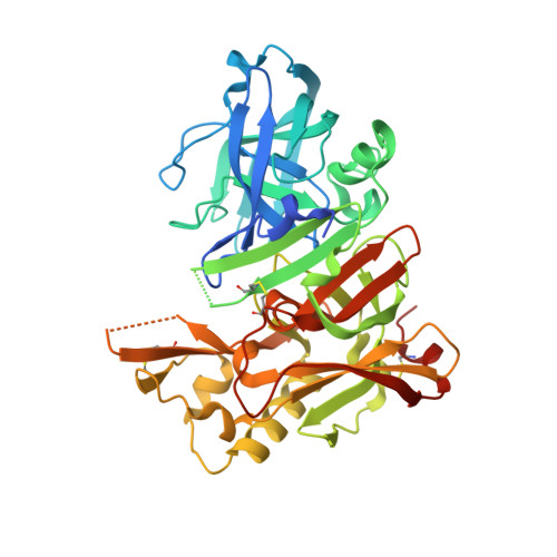
    3EXO

  • PubMed Abstract: 

    A small molecule inhibitor of beta-secretase with a unique binding mode has been developed. Crystallographic determination of the enzyme-inhibitor complex shows the catalytic aspartate residues in the active site are not engaged in inhibitor binding. This unprecedented binding mode in the field of aspartyl protease inhibition is described.


  • Organizational Affiliation

    Department of Medicinal Chemistry, Merck Research Laboratories, Merck & Co, West Point, PA 19486, USA.


Macromolecules
Find similar proteins by:  (by identity cutoff)  |  3D Structure
Entity ID: 1
MoleculeChains Sequence LengthOrganismDetailsImage
Beta-secretase 1413Homo sapiensMutation(s): 2 
Gene Names: BACE1BACEKIAA1149
EC: 3.4.23.46
UniProt & NIH Common Fund Data Resources
Find proteins for P56817 (Homo sapiens)
Explore P56817 
Go to UniProtKB:  P56817
PHAROS:  P56817
GTEx:  ENSG00000186318 
Entity Groups  
Sequence Clusters30% Identity50% Identity70% Identity90% Identity95% Identity100% Identity
UniProt GroupP56817
Sequence Annotations
Expand
  • Reference Sequence
Experimental Data & Validation

Experimental Data

  • Method: X-RAY DIFFRACTION
  • Resolution: 2.10 Å
  • R-Value Free:  0.295 (Depositor), 0.290 (DCC) 
  • R-Value Work:  0.229 (Depositor), 0.230 (DCC) 
  • R-Value Observed: 0.232 (Depositor) 
Space Group: C 2 2 21
Unit Cell:
Length ( Å )Angle ( ˚ )
a = 104.433α = 90
b = 127.923β = 90
c = 76.671γ = 90
Software Package:
Software NamePurpose
DENZOdata reduction
SCALEPACKdata scaling
REFMACrefinement
PDB_EXTRACTdata extraction
StructureStudiodata collection
MOLREPphasing

Structure Validation

View Full Validation Report


Ligand Structure Quality Assessment 

Created with Raphaël 2.3.0Worse 01 BetterLigand structure goodness of fit to experimental dataBest fitted 5MSClick on this verticalbar to view details

Entry History 

Deposition Data

Revision History  (Full details and data files)

  • Version 1.0: 2009-02-03
    Type: Initial release
  • Version 1.1: 2011-07-13
    Changes: Non-polymer description, Version format compliance
  • Version 1.2: 2017-10-25
    Changes: Refinement description
  • Version 1.3: 2021-10-20
    Changes: Database references, Derived calculations
  • Version 1.4: 2023-12-27
    Changes: Data collection
  • Version 1.5: 2024-10-09
    Changes: Structure summary