3GW6 | pdb_00003gw6

Intramolecular Chaperone


Experimental Data Snapshot

  • Method: X-RAY DIFFRACTION
  • Resolution: 2.60 Å
  • R-Value Free: 
    0.255 (Depositor), 0.251 (DCC) 
  • R-Value Work: 
    0.209 (Depositor), 0.205 (DCC) 
  • R-Value Observed: 
    0.211 (Depositor) 

wwPDB Validation 3D Report Full Report

Validation slider image for 3GW6

This is version 1.2 of the entry. See complete history

Literature

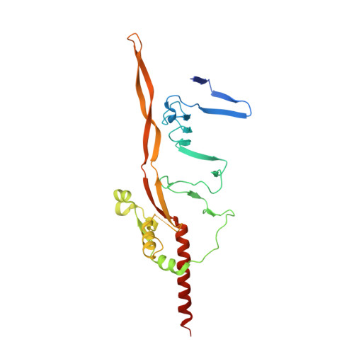
Crystal structure of an intramolecular chaperone mediating triple-beta-helix folding.

Schulz, E.C.Dickmanns, A.Urlaub, H.Schmitt, A.Muhlenhoff, M.Stummeyer, K.Schwarzer, D.Gerardy-Schahn, R.Ficner, R.

(2010) Nat Struct Mol Biol 17: 210-215

  • DOI: https://doi.org/10.1038/nsmb.1746
  • Primary Citation Related Structures: 
    3GUD, 3GW6

  • PubMed Abstract: 

    Protein folding is often mediated by molecular chaperones. Recently, a novel class of intramolecular chaperones has been identified in tailspike proteins of evolutionarily distant viruses, which require a C-terminal chaperone for correct folding. The highly homologous chaperone domains are interchangeable between pre-proteins and release themselves after protein folding. Here we report the crystal structures of two intramolecular chaperone domains in either the released or the pre-cleaved form, revealing the role of the chaperone domain in the formation of a triple-beta-helix fold. Tentacle-like protrusions enclose the polypeptide chains of the pre-protein during the folding process. After the assembly, a sensory mechanism for correctly folded beta-helices triggers a serine-lysine catalytic dyad to autoproteolytically release the mature protein. Sequence analysis shows a conservation of the intramolecular chaperones in functionally unrelated proteins sharing beta-helices as a common structural motif.


  • Organizational Affiliation
    • Abteilung für Molekulare Strukturbiologie, Institut für Mikrobiologie und Genetik, Georg-August-Universität Göttingen, Göttingen, Germany.

Macromolecule Content 

  • Total Structure Weight: 187.38 kDa 
  • Atom Count: 12,617 
  • Modeled Residue Count: 1,531 
  • Deposited Residue Count: 1,650 
  • Unique protein chains: 1

Macromolecules

Find similar proteins by:|  3D Structure
Entity ID: 1
MoleculeChains  Sequence LengthOrganismDetailsImage
Endo-N-acetylneuraminidase275Escherichia phage K1FMutation(s): 0 
Gene Names: sia1717.0
EC: 3.2.1.129
UniProt
Find proteins for Q04830 (Escherichia phage K1F)
Explore Q04830 
Go to UniProtKB:  Q04830
Entity Groups
Sequence Clusters30% Identity50% Identity70% Identity90% Identity95% Identity100% Identity
UniProt GroupQ04830
Sequence Annotations
Expand
Reference Sequence

Small Molecules

Ligands 5 Unique
IDChains Name / Formula / InChI Key2D Diagram3D Interactions
TAM

Query on TAM


Download:Ideal Coordinates CCD File
DA [auth C],
I [auth A],
J [auth A]
TRIS(HYDROXYETHYL)AMINOMETHANE
C7 H17 N O3
GKODZWOPPOTFGA-UHFFFAOYSA-N
PEG

Query on PEG


Download:Ideal Coordinates CCD File
CA [auth C]
EA [auth C]
G [auth A]
GA [auth D]
H [auth A]
CA [auth C],
EA [auth C],
G [auth A],
GA [auth D],
H [auth A],
JA [auth F],
KA [auth F],
LA [auth F],
M [auth B],
MA [auth F],
P [auth E],
Q [auth E],
R [auth E],
S [auth E],
T [auth E],
U [auth E],
V [auth E],
Y [auth C]
DI(HYDROXYETHYL)ETHER
C4 H10 O3
MTHSVFCYNBDYFN-UHFFFAOYSA-N
BR

Query on BR


Download:Ideal Coordinates CCD File
L [auth B]
N [auth B]
NA [auth F]
O [auth B]
W [auth E]
L [auth B],
N [auth B],
NA [auth F],
O [auth B],
W [auth E],
Z [auth C]
BROMIDE ION
Br
CPELXLSAUQHCOX-UHFFFAOYSA-M
CA

Query on CA


Download:Ideal Coordinates CCD File
IA [auth F]CALCIUM ION
Ca
BHPQYMZQTOCNFJ-UHFFFAOYSA-N
CL

Query on CL


Download:Ideal Coordinates CCD File
AA [auth C]
BA [auth C]
FA [auth D]
HA [auth D]
K [auth A]
AA [auth C],
BA [auth C],
FA [auth D],
HA [auth D],
K [auth A],
X [auth E]
CHLORIDE ION
Cl
VEXZGXHMUGYJMC-UHFFFAOYSA-M

Experimental Data & Validation

Experimental Data

  • Method: X-RAY DIFFRACTION
  • Resolution: 2.60 Å
  • R-Value Free:  0.255 (Depositor), 0.251 (DCC) 
  • R-Value Work:  0.209 (Depositor), 0.205 (DCC) 
  • R-Value Observed: 0.211 (Depositor) 
Space Group: P 1
Unit Cell:
Length ( Å )Angle ( ˚ )
a = 62.65α = 81.71
b = 79.95β = 76.53
c = 109.58γ = 87.11
Software Package:
Software NamePurpose
XSCALEdata processing
PHENIXrefinement
PDB_EXTRACTdata extraction
MAR345dtbdata collection
XDSdata reduction
XSCALEdata scaling

Structure Validation

View Full Validation Report


Entry History 

Deposition Data

Revision History  (Full details and data files)

  • Version 1.0: 2010-02-02
    Type: Initial release
  • Version 1.1: 2011-07-13
    Changes: Version format compliance
  • Version 1.2: 2024-02-21
    Changes: Data collection, Database references, Derived calculations