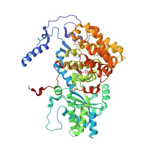
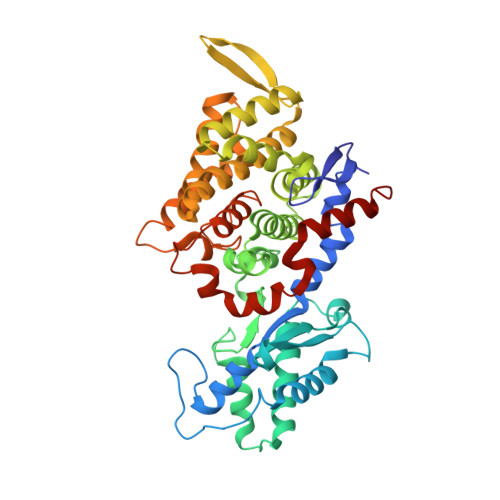
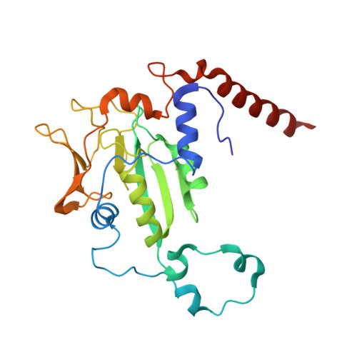
3POT | pdb_00003pot

Structural analysis of a Ni(III)-methyl species in methyl-coenzyme M reductase from Methanothermobacter marburgensis


Experimental Data Snapshot

  • Method: X-RAY DIFFRACTION
  • Resolution: 1.20 Å
  • R-Value Free: 
    0.156 (Depositor), 0.160 (DCC) 
  • R-Value Work: 
    0.132 (Depositor), 0.130 (DCC) 
  • R-Value Observed: 
    0.133 (Depositor) 

Starting Model: experimental
View more details

wwPDB Validation   3D Report Full Report


Ligand Structure Quality Assessment 

Created with Raphaël 2.3.0Worse 01 BetterLigand structure goodness of fit to experimental dataBest fitted F43Click on this verticalbar to view detailsBest fitted TP7Click on this verticalbar to view detailsBest fitted TXZClick on this verticalbar to view details

This is version 1.4 of the entry. See complete history


Literature

Structural Analysis of a Ni-Methyl Species in Methyl-Coenzyme M Reductase from Methanothermobacter marburgensis.

Cedervall, P.E.Dey, M.Li, X.Sarangi, R.Hedman, B.Ragsdale, S.W.Wilmot, C.M.

(2011) J Am Chem Soc 133: 5626-5628

  • DOI: https://doi.org/10.1021/ja110492p
  • Primary Citation of Related Structures:  
    3POT

  • PubMed Abstract: 

    We present the 1.2 Å resolution X-ray crystal structure of a Ni-methyl species that is a proposed catalytic intermediate in methyl-coenzyme M reductase (MCR), the enzyme that catalyzes the biological formation of methane. The methyl group is situated 2.1 Å proximal of the Ni atom of the MCR coenzyme F(430). A rearrangement of the substrate channel has been posited to bring together substrate species, but Ni(III)-methyl formation alone does not lead to any observable structural changes in the channel.


  • Organizational Affiliation

    Department of Biochemistry, Molecular Biology and Biophysics, University of Minnesota, Minneapolis, Minnesota 55455, United States.


Macromolecules
Find similar proteins by:  (by identity cutoff)  |  3D Structure
Entity ID: 1
MoleculeChains Sequence LengthOrganismDetailsImage
Methyl-coenzyme M reductase I subunit alpha
A, D
550Methanothermobacter marburgensisMutation(s): 0 
EC: 2.8.4.1
UniProt
Find proteins for P11558 (Methanothermobacter marburgensis (strain ATCC BAA-927 / DSM 2133 / JCM 14651 / NBRC 100331 / OCM 82 / Marburg))
Explore P11558 
Go to UniProtKB:  P11558
Entity Groups  
Sequence Clusters30% Identity50% Identity70% Identity90% Identity95% Identity100% Identity
UniProt GroupP11558
Sequence Annotations
Expand
  • Reference Sequence
Find similar proteins by:  (by identity cutoff)  |  3D Structure
Entity ID: 2
MoleculeChains Sequence LengthOrganismDetailsImage
Methyl-coenzyme M reductase I subunit beta
B, E
443Methanothermobacter marburgensisMutation(s): 0 
EC: 2.8.4.1
UniProt
Find proteins for P11560 (Methanothermobacter marburgensis (strain ATCC BAA-927 / DSM 2133 / JCM 14651 / NBRC 100331 / OCM 82 / Marburg))
Explore P11560 
Go to UniProtKB:  P11560
Entity Groups  
Sequence Clusters30% Identity50% Identity70% Identity90% Identity95% Identity100% Identity
UniProt GroupP11560
Sequence Annotations
Expand
  • Reference Sequence
Find similar proteins by:  (by identity cutoff)  |  3D Structure
Entity ID: 3
MoleculeChains Sequence LengthOrganismDetailsImage
Methyl-coenzyme M reductase I subunit gamma
C, F
249Methanothermobacter marburgensisMutation(s): 0 
EC: 2.8.4.1
UniProt
Find proteins for P11562 (Methanothermobacter marburgensis (strain ATCC BAA-927 / DSM 2133 / JCM 14651 / NBRC 100331 / OCM 82 / Marburg))
Explore P11562 
Go to UniProtKB:  P11562
Entity Groups  
Sequence Clusters30% Identity50% Identity70% Identity90% Identity95% Identity100% Identity
UniProt GroupP11562
Sequence Annotations
Expand
  • Reference Sequence
Small Molecules
Ligands 8 Unique
IDChains Name / Formula / InChI Key2D Diagram3D Interactions
F43
Query on F43

Download Ideal Coordinates CCD File 
K [auth A],
V [auth D]
FACTOR 430
C42 H51 N6 Ni O13
XLFIRMYGVLUNOY-SXMZNAGASA-M
TP7
Query on TP7

Download Ideal Coordinates CCD File 
N [auth A],
W [auth D]
Coenzyme B
C11 H22 N O7 P S
JBJSVEVEEGOEBZ-SCZZXKLOSA-N
TXZ
Query on TXZ

Download Ideal Coordinates CCD File 
M [auth A],
X [auth D]
O-phosphono-N-(6-sulfanylhexanoyl)-L-threonine
C10 H20 N O7 P S
PRNNQVAYHYUTOD-APPZFPTMSA-N
COM
Query on COM

Download Ideal Coordinates CCD File 
L [auth A],
Y [auth D]
1-THIOETHANESULFONIC ACID
C2 H6 O3 S2
ZNEWHQLOPFWXOF-UHFFFAOYSA-N
06C
Query on 06C

Download Ideal Coordinates CCD File 
O [auth A],
Z [auth D]
Iodomethane
C H3 I
INQOMBQAUSQDDS-UHFFFAOYSA-N
EDO
Query on EDO

Download Ideal Coordinates CCD File 
AA [auth D]1,2-ETHANEDIOL
C2 H6 O2
LYCAIKOWRPUZTN-UHFFFAOYSA-N
K
Query on K

Download Ideal Coordinates CCD File 
P [auth A]POTASSIUM ION
K
NPYPAHLBTDXSSS-UHFFFAOYSA-N
MG
Query on MG

Download Ideal Coordinates CCD File 
BA [auth E]
CA [auth E]
DA [auth F]
G [auth A]
H [auth A]
MAGNESIUM ION
Mg
JLVVSXFLKOJNIY-UHFFFAOYSA-N
Modified Residues  5 Unique
IDChains TypeFormula2D DiagramParent
AGM
Query on AGM
A, D
L-PEPTIDE LINKINGC7 H17 N4 O2ARG
GL3
Query on GL3
A, D
L-PEPTIDE LINKINGC2 H5 N O SGLY
MGN
Query on MGN
A, D
L-PEPTIDE LINKINGC6 H12 N2 O3GLN
MHS
Query on MHS
A, D
L-PEPTIDE LINKINGC7 H11 N3 O2HIS
SMC
Query on SMC
A, D
L-PEPTIDE LINKINGC4 H9 N O2 SCYS
Experimental Data & Validation

Experimental Data

  • Method: X-RAY DIFFRACTION
  • Resolution: 1.20 Å
  • R-Value Free:  0.156 (Depositor), 0.160 (DCC) 
  • R-Value Work:  0.132 (Depositor), 0.130 (DCC) 
  • R-Value Observed: 0.133 (Depositor) 
Space Group: P 1 21 1
Unit Cell:
Length ( Å )Angle ( ˚ )
a = 81.886α = 90
b = 118.165β = 91.94
c = 122.388γ = 90
Software Package:
Software NamePurpose
REFMACrefinement
HKL-2000data reduction
HKL-2000data scaling
REFMACphasing

Structure Validation

View Full Validation Report


Ligand Structure Quality Assessment 

Created with Raphaël 2.3.0Worse 01 BetterLigand structure goodness of fit to experimental dataBest fitted F43Click on this verticalbar to view detailsBest fitted TP7Click on this verticalbar to view detailsBest fitted TXZClick on this verticalbar to view details

Entry History 

Deposition Data

Revision History  (Full details and data files)

  • Version 1.0: 2011-04-13
    Type: Initial release
  • Version 1.1: 2011-07-13
    Changes: Version format compliance
  • Version 1.2: 2015-05-20
    Changes: Non-polymer description
  • Version 1.3: 2017-03-29
    Changes: Non-polymer description
  • Version 1.4: 2023-09-06
    Changes: Data collection, Database references, Derived calculations, Refinement description