4XC6

Isobutyryl-CoA mutase fused with bound adenosylcobalamin, GDP, and Mg (holo-IcmF/GDP)


Experimental Data Snapshot

  • Method: X-RAY DIFFRACTION
  • Resolution: 3.35 Å
  • R-Value Free: 
    0.198 (Depositor), 0.200 (DCC) 
  • R-Value Work: 
    0.180 (Depositor), 0.180 (DCC) 
  • R-Value Observed: 
    0.181 (Depositor) 

wwPDB Validation   3D Report Full Report


Ligand Structure Quality Assessment 

Created with Raphaël 2.3.0Worse 01 BetterLigand structure goodness of fit to experimental dataBest fitted B12Click on this verticalbar to view detailsBest fitted GDPClick on this verticalbar to view detailsBest fitted 5ADClick on this verticalbar to view details

This is version 1.4 of the entry. See complete history


Literature

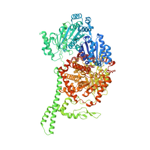
Visualization of a radical B12 enzyme with its G-protein chaperone.

Jost, M.Cracan, V.Hubbard, P.A.Banerjee, R.Drennan, C.L.

(2015) Proc Natl Acad Sci U S A 112: 2419-2424

  • DOI: https://doi.org/10.1073/pnas.1419582112
  • Primary Citation of Related Structures:  
    4XC6, 4XC7, 4XC8

  • PubMed Abstract: 

    G-protein metallochaperones ensure fidelity during cofactor assembly for a variety of metalloproteins, including adenosylcobalamin (AdoCbl)-dependent methylmalonyl-CoA mutase and hydrogenase, and thus have both medical and biofuel development applications. Here, we present crystal structures of IcmF, a natural fusion protein of AdoCbl-dependent isobutyryl-CoA mutase and its corresponding G-protein chaperone, which reveal the molecular architecture of a G-protein metallochaperone in complex with its target protein. These structures show that conserved G-protein elements become ordered upon target protein association, creating the molecular pathways that both sense and report on the cofactor loading state. Structures determined of both apo- and holo-forms of IcmF depict both open and closed enzyme states, in which the cofactor-binding domain is alternatively positioned for cofactor loading and for catalysis. Notably, the G protein moves as a unit with the cofactor-binding domain, providing a visualization of how a chaperone assists in the sequestering of a precious cofactor inside an enzyme active site.


  • Organizational Affiliation

    Department of Chemistry and.


Macromolecules
Find similar proteins by:  (by identity cutoff)  |  3D Structure
Entity ID: 1
MoleculeChains Sequence LengthOrganismDetailsImage
Isobutyryl-CoA mutase fused
A, B
1,113Cupriavidus metallidurans CH34Mutation(s): 0 
Gene Names: IcmFRmet_0210
EC: 5.4.99.13 (PDB Primary Data), 3.6.5 (UniProt)
UniProt
Find proteins for Q1LRY0 (Cupriavidus metallidurans (strain ATCC 43123 / DSM 2839 / NBRC 102507 / CH34))
Explore Q1LRY0 
Go to UniProtKB:  Q1LRY0
Entity Groups  
Sequence Clusters30% Identity50% Identity70% Identity90% Identity95% Identity100% Identity
UniProt GroupQ1LRY0
Sequence Annotations
Expand
  • Reference Sequence
Small Molecules
Ligands 5 Unique
IDChains Name / Formula / InChI Key2D Diagram3D Interactions
B12
Query on B12

Download Ideal Coordinates CCD File 
C [auth A],
I [auth B]
COBALAMIN
C62 H89 Co N13 O14 P
LKVIQTCSMMVGFU-DWSMJLPVSA-N
GDP
Query on GDP

Download Ideal Coordinates CCD File 
E [auth A],
J [auth B]
GUANOSINE-5'-DIPHOSPHATE
C10 H15 N5 O11 P2
QGWNDRXFNXRZMB-UUOKFMHZSA-N
5AD
Query on 5AD

Download Ideal Coordinates CCD File 
D [auth A]5'-DEOXYADENOSINE
C10 H13 N5 O3
XGYIMTFOTBMPFP-KQYNXXCUSA-N
ACT
Query on ACT

Download Ideal Coordinates CCD File 
H [auth A]ACETATE ION
C2 H3 O2
QTBSBXVTEAMEQO-UHFFFAOYSA-M
MG
Query on MG

Download Ideal Coordinates CCD File 
F [auth A],
G [auth A],
K [auth B],
L [auth B]
MAGNESIUM ION
Mg
JLVVSXFLKOJNIY-UHFFFAOYSA-N
Experimental Data & Validation

Experimental Data

  • Method: X-RAY DIFFRACTION
  • Resolution: 3.35 Å
  • R-Value Free:  0.198 (Depositor), 0.200 (DCC) 
  • R-Value Work:  0.180 (Depositor), 0.180 (DCC) 
  • R-Value Observed: 0.181 (Depositor) 
Space Group: H 3 2
Unit Cell:
Length ( Å )Angle ( ˚ )
a = 318.76α = 90
b = 318.76β = 90
c = 343.52γ = 120
Software Package:
Software NamePurpose
PHENIXrefinement
XDSdata reduction
XSCALEdata scaling
SHELXphasing
SHARPphasing
Cootmodel building

Structure Validation

View Full Validation Report


Ligand Structure Quality Assessment 

Created with Raphaël 2.3.0Worse 01 BetterLigand structure goodness of fit to experimental dataBest fitted B12Click on this verticalbar to view detailsBest fitted GDPClick on this verticalbar to view detailsBest fitted 5ADClick on this verticalbar to view details

Entry History & Funding Information

Deposition Data


Funding OrganizationLocationGrant Number
National Institutes of Health/National Institute of General Medical Sciences (NIH/NIGMS)United StatesGM069857
Howard Hughes Medical Institute (HHMI)United States--

Revision History  (Full details and data files)

  • Version 1.0: 2015-02-11
    Type: Initial release
  • Version 1.1: 2015-03-04
    Changes: Database references
  • Version 1.2: 2017-09-13
    Changes: Author supporting evidence, Database references, Derived calculations, Source and taxonomy
  • Version 1.3: 2019-11-20
    Changes: Author supporting evidence
  • Version 1.4: 2024-02-28
    Changes: Data collection, Database references, Derived calculations