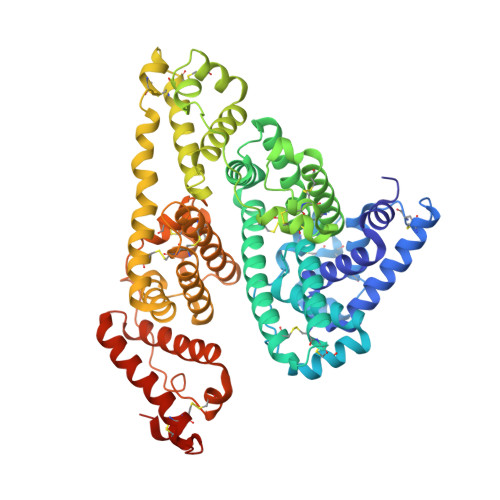
5YOQ

Human serum albumin complexed with sodium 4-phenylbutyrate


Experimental Data Snapshot

  • Method: X-RAY DIFFRACTION
  • Resolution: 2.65 Å
  • R-Value Free: 0.256 
  • R-Value Work: 0.235 
  • R-Value Observed: 0.236 

Starting Model: experimental
View more details

wwPDB Validation   3D Report Full Report


Ligand Structure Quality Assessment 


This is version 1.3 of the entry. See complete history


Literature

Crystal structure analysis of human serum albumin complexed with sodium 4-phenylbutyrate.

Kawai, A.Yamasaki, K.Enokida, T.Miyamoto, S.Otagiri, M.

(2018) Biochem Biophys Rep 13: 78-82

  • DOI: https://doi.org/10.1016/j.bbrep.2018.01.006
  • Primary Citation of Related Structures:  
    5YOQ

  • PubMed Abstract: 

    Sodium 4-phenylbutyrate (PB) is an orphan drug for the treatment of urea cycle disorders. It also inhibits the development of endoplasmic reticulum stress, the action of histone deacetylases and as a regulator of the hepatocanalicular transporter. PB is generally considered to have the potential for use in the treatment of the diseases such as cancer, neurodegenerative diseases and metabolic diseases. In a previous study, we reported that PB is primarily bound to human serum albumin (HSA) in plasma and its binding site is drug site 2. However, details of the binding mode of PB to HSA remain unknown. To address this issue, we examined the crystal structure of HSA with PB bound to it. The structure of the HSA-PB complex indicates that the binding mode of PB to HSA is quite similar to that for octanoate or drugs that bind to drug site 2, as opposed to that for other medium-chain length of fatty acids. These findings provide useful basic information related to drug-HSA interactions. Moreover, the information presented herein is valuable in terms of providing safe and efficient treatment and diagnosis in clinical settings.


  • Organizational Affiliation

    Faculty of Pharmaceutical Sciences, Sojo University, 4-22-1 Ikeda, Nishi-ku, Kumamoto 860-0082, Japan.


Macromolecules
Find similar proteins by:  (by identity cutoff)  |  3D Structure
Entity ID: 1
MoleculeChains Sequence LengthOrganismDetailsImage
Serum albumin
A, B
585Homo sapiensMutation(s): 0 
Gene Names: ALBGIG20GIG42PRO0903PRO1708PRO2044PRO2619PRO2675UNQ696/PRO1341
UniProt & NIH Common Fund Data Resources
Find proteins for P02768 (Homo sapiens)
Explore P02768 
Go to UniProtKB:  P02768
PHAROS:  P02768
GTEx:  ENSG00000163631 
Entity Groups  
Sequence Clusters30% Identity50% Identity70% Identity90% Identity95% Identity100% Identity
UniProt GroupP02768
Sequence Annotations
Expand
  • Reference Sequence
Experimental Data & Validation

Experimental Data

  • Method: X-RAY DIFFRACTION
  • Resolution: 2.65 Å
  • R-Value Free: 0.256 
  • R-Value Work: 0.235 
  • R-Value Observed: 0.236 
  • Space Group: P 1 21 1
Unit Cell:
Length ( Å )Angle ( ˚ )
a = 58.465α = 90
b = 181.913β = 105.16
c = 59.474γ = 90
Software Package:
Software NamePurpose
PHENIXrefinement
HKL-2000data reduction
HKL-2000data scaling
MOLREPphasing

Structure Validation

View Full Validation Report


Ligand Structure Quality Assessment 


Entry History 

Deposition Data

Revision History  (Full details and data files)

  • Version 1.0: 2018-02-07
    Type: Initial release
  • Version 1.1: 2018-02-28
    Changes: Database references
  • Version 1.2: 2023-11-22
    Changes: Data collection, Database references, Refinement description
  • Version 1.3: 2024-11-13
    Changes: Structure summary